. 2022 Feb 28;11(3):475.
doi: 10.3390/antiox11030475.

Polyphenol Rich Forsythia suspensa Extract Alleviates DSS-Induced Ulcerative Colitis in Mice through the Nrf2-NLRP3 Pathway

Affiliations

Polyphenol Rich Forsythia suspensa Extract Alleviates DSS-Induced Ulcerative Colitis in Mice through the Nrf2-NLRP3 Pathway

Limin Chao et al. Antioxidants (Basel). .

Abstract

This study systematically evaluated the effect of Forsythia suspensa extract on dextran sodium sulfate (DSS)-induced ulcerative colitis (UC) and determined its mechanism of action. The results showed that Forsythia suspensa extract significantly inhibited DSS-induced UC in mice. In vivo mechanistic studies revealed that Forsythia suspensa extract relieved the symptoms of colitis by enhancing antioxidant activity and inhibiting pyroptosis. Further in vitro experiments on the mechanism of Forsythia suspensa showed that it reduced the level of reactive oxygen species (ROS) in J774A.1 cells. We found that Forsythia suspensa extract enhanced cellular antioxidation activity and inhibited pyroptosis. After silencing NLRP3, it was found to play an important role in pyroptosis. In addition, after Nrf2 was silenced, the inhibitory effect of Forsythia suspensa extract on cell pyroptosis was eliminated, indicating an interaction between Nrf2 and NLRP3. Metabonomics revealed that Forsythia suspensa extract significantly improved metabolic function in colitis mice by reversing the abnormal changes in the levels of 9 metabolites. The main metabolic pathways involved were glutathione metabolism, aminoacyl-tRNA biosynthesis and linoleic acid metabolism. In conclusion, we found that Forsythia suspensa extract significantly alleviated DSS-induced UC injury through the Nrf2-NLRP3 pathway and relieved metabolic dysfunction.

Keywords: Forsythia suspensa; antioxidant; metabolomics; pyroptosis; ulcerative colitis.


Conflict of interest statement

The authors declare no conflict of interest.

Figures

Figure 1
Figure 1
Forsythia suspensa extract alleviates the symptoms of UC in mice. (A) Average water consumption during modelling. (B) Average weight of the mice (n = 10). (C) DAI score. (D) Representative pictures of the colon. (E) Average colon length (n = 10). (F) Measurement of the level of IL-1β in the serum with an ELISA kit. (G) Pathological examination of mouse colon tissue (×200). (H) Histological score of the colon. Data represent at least three independent experiments. The results are shown as the mean ± SEM. # p < 0.05, and ## p < 0.01 compared with the control group; * p < 0.05, and ** p < 0.01 compared with the model group; #** means # and **.
Figure 2
Figure 2
The effect of Forsythia suspensa extract on the levels of MDA, SOD and MPO in mice. (A) MDA level in serum. (B) SOD level in serum. (C) MPO activity in colon tissue. Data represent at least three independent experiments. The results are shown as the mean ± SEM. # p < 0.05, and ## p < 0.01 compared with the control group; * p < 0.05, and ** p < 0.01 compared with the model group; #** means # and **; ##** means ## and **.
Figure 3
Figure 3
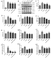
Forsythia suspensa extract enhances the antioxidant activity in mouse colon tissue. (AE) Western blot analysis of cytoplasm for Nrf2, HO-1, NQO1 and Keap1. (FI) qRT–PCR detection of Nrf2, HO-1, NQO1 and Keap1 gene expression levels. (J) Western blot analysis of the level of nuclear Nrf2. The results are shown as the mean ± SEM. # p < 0.05, and ## p < 0.01 compared with the control group; * p < 0.05, and ** p < 0.01 compared with the model group; #** means # and **; ##** means ## and **.
Figure 4
Figure 4
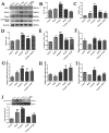
Forsythia suspensa extract may alleviate colitis by inhibiting pyroptosis. (AF) Western blot analysis of ASC, Caspase-1, IL-1β, GSDMD and NLRP3. (GK) qRT–PCR detection of ASC, Caspase-1, IL-1β, GSDMD and NLRP3 gene expression levels. The results are shown as the mean ± SEM. # p < 0.05, and ## p < 0.01 compared with the control group; * p < 0.05, and ** p < 0.01 com-pared with the model group; #** means # and **; ##** means ## and **.
Figure 5
Figure 5
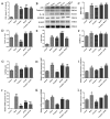
Forsythia suspensa extract can reduce the protein and gene expression of pyroptosis in J774A.1 cells. (A) J774A.1 cells were first treated with 1 μg/mL LPS and different concentrations of Forsythia suspensa extract for 12 h, and then stimulated with 5 mM ATP for 1 h. The kit detects the level of LDH release. (BG) Western blot analysis of ASC, Caspase-1, IL-1β, GSDMD and NLRP3. (HL) qRT–PCR detection of ASC, Caspase-1, IL-1β, GSDMD and NLRP3 gene expression levels. Data represent at least three independent experiments. The results are shown as the mean ± SEM. # p < 0.05, and ## p < 0.01 compared with the control group; * p < 0.05, and ** p < 0.01 compared with the model group; #** means # and **; ##** means ## and **.
Figure 6
Figure 6
Forsythia suspensa extract enhances the antioxidant activity J774A.1 cells. (AE) Western blot analysis of HO-1, NQO1, and cytoplasm for Nrf2 and Keap1. (FI) qRT–PCR detection of HO-1, NQO1, Nrf2 and Keap1 gene expression levels. (J) Western blot analysis of the level of nuclear Nrf2. The results are shown as the mean ± SEM. # p < 0.05, and ## p < 0.01 compared with the control group; * p < 0.05, and ** p < 0.01 compared with the model group; #** means # and **; ##** means ## and **.
Figure 7
Figure 7
Forsythia suspensa extract significantly reduces the level of ROS in J774A.1 cells. (A) Control group. (B) Model group. (C) 0.5 mg/mL Forsythia suspensa extract. (D) 1 mg/mL Forsythia suspensa extract. (E) 2 mg/mL Forsythia suspensa extract; (F) Statistical analysis of ROS levels in J774A.1 cells. Data represent at least three independent experiments. The results are shown as the mean ± SEM. ## p < 0.01 compared with the control group;** p < 0.01 compared with the model group; #** means # and **; ##** means ## and **.
Figure 8
Figure 8
Forsythia suspensa extract may block the occurrence of pyroptosis by inhibiting the activation of NLRP3. (A) LDH levels in J774A.1 cells. (BG) Western blot analysis of ASC, Caspase-1, IL-1β, GSDMD and NLRP3. (HL) qRT–PCR detection of ASC, Caspase-1, IL-1β, GSDMD and NLRP3 gene expression levels. Data represent at least three independent experiments. The results are shown as the mean ± SEM.# p < 0.05, and ## p < 0.01 compared with the control group; * p < 0.05, and ** p < 0.01 compared with the model group; #** means # and **; ##** means ## and **.
Figure 9
Figure 9
The inhibitor ML385 reverses the antioxidant effect of Forsythia suspensa extract. (AE) Western blot analysis of HO-1, NQO1, Keap1 and cytoplasm for Nrf2. (FI) qRT–PCR detection of HO-1, NQO1, Keap1 and Nrf2 gene expression levels. (J) Western blot detects the level of nuclear Nrf2. The results are shown as the mean ± SEM. # p < 0.05, and ## p < 0.01 compared with the control group; * p < 0.05, and ** p < 0.01 compared with the model group; #** means # and **; ##** means ## and **.
Figure 10
Figure 10
The effect of Forsythia suspensa extract in inhibiting pyroptosis may be related to its antioxidant effect. (A) LDH levels in J774A.1 cells. (BG) Western blot analysis of ASC, Caspase-1, IL-1β, GSDMD and NLRP3. (HL) qRT–PCR detection of ASC, Caspase-1, IL-1β, GSDMD and NLRP3 gene expression levels. Data represent at least three independent experiments. The results are shown as the mean ± SEM. # p < 0.05, and ## p < 0.01 compared with the control group; * p < 0.05, and ** p < 0.01 compared with the model group; ##* means ## and *; ##** means ## and **.
Figure 11
Figure 11
The OPLS-DA scores of the control, model, L-dose, M-dose and H-dose groups are based on the serum metabolite curve in positive and negative ion modes. (A) Positive ion mode; (B) Negative ion mode.
Figure 12
Figure 12
Serum levels of nine metabolites. (A) Glutathione, (B) 13-L-hydroperoxylinoleic acid, (C) 13-oxoODE, (D) L-aspartic acid, (E) L-leucine, (F) L-lysine, (G) L-isoleucine, (H) L-tryptophan and (I) L-methionine. Data represent at least three independent experiments. The results are shown as the mean ± SEM. # p < 0.05, and ## p < 0.01 compared with the control group; * p < 0.05, and ** p < 0.01 compared with the model group; #** means # and **; ##** means ## and **.
Figure 13
Figure 13
Metabolic pathway analysis based on the potential biomarkers. 1, glutathione metabolism; 2, aminoacyl-tRNA biosynthesis; 3, linoleic acid metabolism.

Similar articles

Cited by

References

    1. Ungaro R., Mehandru S., Allen P.B., Peyrin-Biroulet L., Colombel J.F. Ulcerative colitis. Lancet. 2017;389:1756–1770. doi: 10.1016/S0140-6736(16)32126-2. - DOI - PMC - PubMed
    1. Zhang Z., Shen P., Xie W., Cao H., Liu J., Cao Y., Zhang N. Pingwei San ameliorates dextran sulfate sodium-induced chronic colitis in mice. J. Ethnopharmacol. 2019;236:91–99. doi: 10.1016/j.jep.2019.01.043. - DOI - PubMed
    1. Wang X., Fan F., Cao Q. Modified Pulsatilla decoction attenuates oxazolone-induced colitis in mice through suppression of inflammation and epithelial barrier disruption. Mol. Med. Rep. 2016;14:1173–1179. doi: 10.3892/mmr.2016.5358. - DOI - PMC - PubMed
    1. de Souza H. Etiopathogenesis of inflammatory bowel disease: Today and tomorrow. Curr. Opin. Gastroenterol. 2017;33:222–229. doi: 10.1097/MOG.0000000000000364. - DOI - PubMed
    1. Abderrazak A., Syrovets T., Couchie D., El H.K., Friguet B., Simmet T., Rouis M. NLRP3 inflammasome: From a danger signal sensor to a regulatory node of oxidative stress and inflammatory diseases. Redox Biol. 2015;4:296–307. doi: 10.1016/j.redox.2015.01.008. - DOI - PMC - PubMed

LinkOut - more resources