Review
. 2022 Mar 4;11(3):501.
doi: 10.3390/antiox11030501.

Superoxide Radicals in the Execution of Cell Death

Affiliations
Review

Superoxide Radicals in the Execution of Cell Death

Junichi Fujii et al. Antioxidants (Basel). .

Abstract

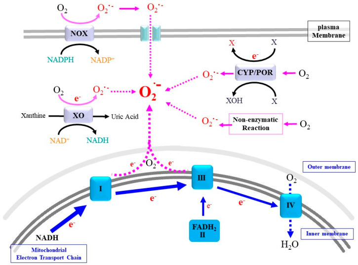
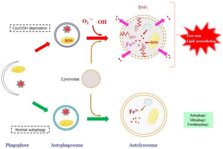
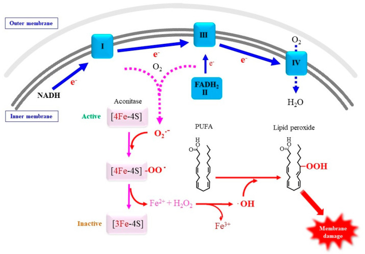
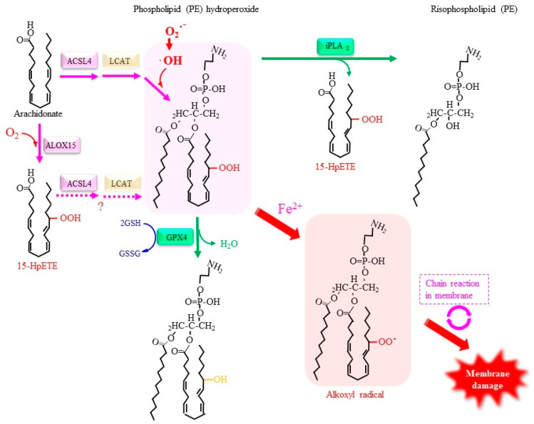
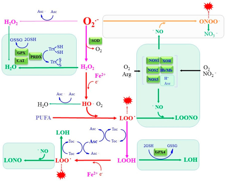
Superoxide is a primary oxygen radical that is produced when an oxygen molecule receives one electron. Superoxide dismutase (SOD) plays a primary role in the cellular defense against an oxidative insult by ROS. However, the resulting hydrogen peroxide is still reactive and, in the presence of free ferrous iron, may produce hydroxyl radicals and exacerbate diseases. Polyunsaturated fatty acids are the preferred target of hydroxyl radicals. Ferroptosis, a type of necrotic cell death induced by lipid peroxides in the presence of free iron, has attracted considerable interest because of its role in the pathogenesis of many diseases. Radical electrons, namely those released from mitochondrial electron transfer complexes, and those produced by enzymatic reactions, such as lipoxygenases, appear to cause lipid peroxidation. While GPX4 is the most potent anti-ferroptotic enzyme that is known to reduce lipid peroxides to alcohols, other antioxidative enzymes are also indirectly involved in protection against ferroptosis. Moreover, several low molecular weight compounds that include α-tocopherol, ascorbate, and nitric oxide also efficiently neutralize radical electrons, thereby suppressing ferroptosis. The removal of radical electrons in the early stages is of primary importance in protecting against ferroptosis and other diseases that are related to oxidative stress.

Keywords: ferroptosis; nitric oxide; radical electron; superoxide.


Conflict of interest statement

The authors declare no conflict of interest.

Figures

Figure 1
Figure 1
Representative electron sources of superoxide. While mitochondrial ETC is the major source for superoxide (O2.−), many enzymes, such as NADPH oxidase (NOX), xanthine oxidase (XO), and cytochrome P450 (CYP)/cytochrome P450 reductase (POR), convert molecular oxygen to superoxide either as a main product or as a byproduct during oxidation of a variety of compounds (X), such as benzene compounds, drugs, and steroid hormones. Superoxide is also produced non-enzymatically.
Figure 2
Figure 2
A role of ferritinophagy in releasing free iron. In normal ferritinophagy, free iron is released from ferritin but remains in the autolysosomes, which does not cause deteriorating effects as schematized at the bottom. Under ferroptotic conditions, the produced ROS causes lipid peroxidation. The presence of peroxidized lipids and free iron would make autolysosome membrane vulnerable, leading to the destruction of the vesicular structure.
Figure 3
Figure 3
Proposed role of superoxide in mitochondrial membrane damage. Superoxide that is released from the ETC under ferroptotic stimuli may become attached to 4Fe-4S clusters in proteins, notably aconitase, which results in the release of ferrous iron and hydrogen peroxide. PUFA in mitochondrial membrane undergoes peroxidation by ROS and consequently destructs mitochondrial membrane integrity.
Figure 4
Figure 4
ALOX15 is a representative enzyme that catalyzes the peroxidation of arachidonate. ACSL4 preferentially acts on arachidonate derivatives and forms acyl-coenzyme A, which is then used to construct phospholipids by LCAT. Lipid peroxides in phospholipid bilayers react with free iron and result in the production of alkoxyl radicals, which enhances lipid peroxidation, ultimately leading to the destruction of the membrane structure. iPLA2 preferentially removes an acyl group at the sn-2 position where an unsaturated fatty acid is conjugated such that 15-HpETE can be excised by the action of iPLA2, which leads to the membrane structure being protected. Conversely, GPX4 interrupts the fatal chain reaction by reducing 15-HpETE to 15-HETE in the form of phospholipid hydroperoxide.
Figure 5
Figure 5
The interaction between ROS/lipid radicals with antioxidants in vivo. Detoxification reactions performed by representative antioxidative enzymes and low molecular weight antioxidants are presented in green. In addition to antioxidative enzymes, Toc, Asc, and NO can terminate radical chain reactions, which result in the suppression of ferroptosis.

Similar articles

Cited by

References

    1. Fridovich I. Superoxide radical and superoxide dismutases. Annu. Rev. Biochem. 1995;64:97–112. doi: 10.1146/annurev.bi.64.070195.000525. - DOI - PubMed
    1. Rhee S.G. Cell signaling. H2O2, a necessary evil for cell signaling. Science. 2006;312:1882–1883. doi: 10.1126/science.1130481. - DOI - PubMed
    1. Finkel T. Signal transduction by reactive oxygen species. J. Cell Biol. 2011;194:7–15. doi: 10.1083/jcb.201102095. - DOI - PMC - PubMed
    1. Lennicke C., Cochemé H.M. Redox metabolism: ROS as specific molecular regulators of cell signaling and function. Mol. Cell. 2021;81:3691–3707. doi: 10.1016/j.molcel.2021.08.018. - DOI - PubMed
    1. Janssen-Heininger Y.M., Mossman B.T., Heintz N.H., Forman H.J., Kalyanaraman B., Finkel T., Stamler J.S., Rhee S.G., van der Vliet A. Redox-based regulation of signal transduction: Principles, pitfalls, and promises. Free Radic. Biol. Med. 2008;45:1–17. doi: 10.1016/j.freeradbiomed.2008.03.011. - DOI - PMC - PubMed

Grants and funding

LinkOut - more resources