. 2022 Mar 10;11(3):530.
doi: 10.3390/antiox11030530.

Piperine Attenuates Lithocholic Acid-Stimulated Interleukin-8 by Suppressing Src/EGFR and Reactive Oxygen Species in Human Colorectal Cancer Cells

Affiliations

Piperine Attenuates Lithocholic Acid-Stimulated Interleukin-8 by Suppressing Src/EGFR and Reactive Oxygen Species in Human Colorectal Cancer Cells

Shinan Li et al. Antioxidants (Basel). .

Abstract

Piperine, a natural alkaloidal pungent product present in pepper plants, possesses the properties of anti-inflammatory and anti-metastasis. Lithocholic acid is a monohydroxy-5beta-cholanic acid with an alpha-hydroxy substituent at position 3; it is a secondary bile acid that plays a pivotal role in fat absorption, and has been discovered to mediate colorectal cancer (CRC) cell invasion and migration. However, the effect of piperine on angiogenesis has been poorly investigated. In the current study, we examined the role of piperine on LCA-stimulated angiogenesis by measuring interleukin-8 (IL-8) expression; moreover, we revealed the potential molecular mechanisms in CRC cells. Here, we showed that piperine inhibited LCA-stimulated endothelial EA.hy926 cell angiogenesis in a conditioned medium obtained from colorectal HCT-116 cells. Experiments with an IL-8 neutralizer showed that IL-8 present in the conditioned medium was the major angiogenic factor. Piperine inhibited LCA-stimulated ERK1/2 and AKT via the Src/EGFR-driven ROS signaling pathway in the colorectal cell line (HCT-116). Through mutagenesis and inhibitory studies, we revealed that ERK1/2 acted as an upstream signaling molecule in AP-1 activation, and AKT acted as an upstream signaling molecule in NF-κB activation, which in turn attenuated IL-8 expression. Taken together, we demonstrated that piperine blocked LCA-stimulated IL-8 expression by suppressing Src and EGFR in human CRC HCT-116 cells, thus remarkably attenuating endothelial EA.hy926 cell tube formation.

Keywords: colorectal cancer; interleukin-8; piperine; reactive oxygen species; tumor microenvironment.


Conflict of interest statement

The authors declare no conflict of interest regarding the publication of this paper. Authors Thi Thinh Nguyen and Trong Thuan Ung are from Nanogen Pharmaceutical Biotechnology Joint Stock Company, the company had no role in the design of the study; in the collection, analyses, or interpretation of data; in the writing of the manuscript, or in the decision to publish the results.

Figures

Figure 1
Figure 1
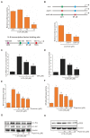
Piperine Inhibits LCA-Induced IL-8 Expression in HCT-116 Cells. (A) Four different human CRC cell lines were incubated with 30 μM lithocholic acid (LCA) for 4 h, followed by mRNA extraction and RT-qPCR, to determine IL-8 expression level. HCT-116 cells were incubated with 30 μΜ LCA for 0–4 h (B) or with 0–30 μM LCA for 4 h (C). (D) HCT-116 cells were transiently transfected with 500 ng pGL2-IL-8 promoter–reporter construct. These transfected cells were incubated with 30 μΜ LCA in a dose-dependent manner for 4 h, and the luciferase activity was measured using a luminometer. (E) Three different human CRC cell lines were pretreated with 30 μΜ piperine and incubated with 30 μΜ LCA for 4 h, followed by mRNA extraction and RT-qPCR to determine IL-8 expression level. (F) HCT-116 cells were pretreated with piperine (10, 30, and 50 μΜ) and incubated with 30 μΜ LCA for 4 h, followed by mRNA extraction and RT-qPCR to determine IL-8 expression level. (G) HCT-116 cells were transiently transfected with 500 ng pGL2-IL-8 promoter–reporter construct. These transfected cells were pretreated with piperine (10, 30, and 50 μΜ) and incubated with 30 μΜ LCA for 4 h, and the luciferase activity was measured using a luminometer. (H) HCT-116 cells were pretreated with piperine (10, 30, and 50 μΜ) and incubated with 30 μΜ LCA for 24 h, followed by ELISA assay to determine the IL-8 secretion level. Data represent the mean ± standard error of the mean (SEM) from three experimental trials. * p < 0.05 versus control; # p < 0.05 versus LCA.
Figure 2
Figure 2
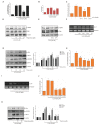
Piperine Inhibits LCA-Induced IL-8 by Suppressing the Transcriptional Activities of AP-1 and NF-κB in HCT-116 Cells. (A) The IL-8 promoter was sequentially deleted in the 5′-flanking region, and the promoter luciferase construct was transiently transfected into HCT-116 cells. These transfected cells were incubated with 30 μΜ LCA for 4 h, and the luciferase activity was measured using a luminometer. Data represent the mean ± SEM from three experimental trials. * p < 0.05 versus –133; # p < 0.05 versus –98. (B) The IL-8 promoter was mutated at the NF-κB and AP-1 binding sites (mNF-κB and mAP-1) and the promoter luciferase construct was transiently transfected into HCT-116 cells. The transfected cells were incubated with 30 μΜ LCA for 4 h, and the luciferase activity was measured using a luminometer. Data represent the mean ± SEM from three experimental trials. * p < 0.05 versus LCA only. HCT-116 cells were pretreated with SR-11302 (SR (C)), BAY-11-7082 (BAY, 20 μM (D)), or SN50 (50 μg/mL (D)) for 1 h and incubated with 30 μΜ LCA for 4 h, followed by mRNA extraction and RT-qPCR to determine IL-8 secretion level. HCT-116 cells were transiently transfected with AP-1 luciferase reporter construct (E) or NF-κB luciferase reporter construct (F). These transfected cells were pretreated with piperine (10, 30, and 50 μΜ) and incubated with 30 μΜ LCA for 4 h, and the luciferase activity was measured using a luminometer. Data represent the mean ± SEM from three experimental trials. * p < 0.05 versus control; # p < 0.05 versus IL-8. (G) HCT-116 cells were pretreated with piperine (10, 30, and 50 μΜ) and incubated with 30 μΜ LCA for 4 h; cell lysates were analyzed for phosphorylated c-Fos and c-Jun level using western blotting. (H) HCT-116 cells were pretreated with piperine (10, 30, and 50 μΜ) and incubated with 30 μΜ LCA for 4 h, cell lysates were analyzed for phosphorylated p65 level using western blotting.
Figure 3
Figure 3
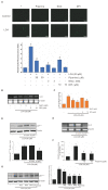
Piperine Inhibits LCA-Induced IL-8 Expression by Suppressing the Activation of ERK1/2, Src, AKT, and EGFR Signaling Pathways. (A) HCT-116 cells were pretreated with 30 μM concentration of SB-203580 (SB), 30 μM concentration of PD-98059 (PD), and 30 μM concentration of JNKi for 1 h, and incubated with 30 μM concentration of LCA for 4 h. Subsequently, the IL-8 mRNA level was measured through RT-qPCR. (B) HCT-116 cells pretreated with 30 μM SB, 30 μM PD, and 30 μM JNKi for 1 h were incubated with 30 μM LCA for 24 h followed by ELISA assay to determine the IL-8 expression level. (C) HCT-116 cells were transiently transfected with dominant-negative mutants of MEK-1 (K97 M), JNK (TAM67), or mutant p38 MAPK (mP38), and co-transfected with pGL2-IL-8. After incubation with 30 μM LCA for 4 h, the luciferase activity was measured using a luminometer. (D) HCT-116 cells pretreated with 30 μM SB, 30 μM PD, or 30 μM JNKi for 1 h were incubated with 30 μM LCA for 4 h, and cell lysates were analyzed for the phosphorylated and total ERK level using western blotting. (E) HCT-116 cells pretreated with 5 mM NAC or 10 μM DPI for 1 h were incubated with 30 μM LCA for 4 h, and cell lysates were analyzed for the phosphorylated and total ERK level using western blotting. (F) HCT-116 cells were incubated with 30 μM LCA for 0–60 min, and cell lysates were analyzed for levels of phosphorylated Src, AKT, and EGFR using western blotting. (G) HCT-116 cells pretreated with 10 μM AG1478 (AG), 10 μM PP1, 10 μM PP2, and 20 μM LY-294002 (LY) for 1 h were incubated with 30 μM LCA for 4 h. Subsequently, the IL-8 mRNA level was measured using RT-PCR. (H) HCT-116 cells pretreated with 10 μM AG, 10 μM PP1, 10 μM PP2, and 20 μM LY for 1 h were incubated with 30 μM LCA for 4 h, and the luciferase activity was measured using a luminometer. (I) Cells transfected with si-Con, si-Src, si-EGFR, and si-AKT were incubated with 30 μM LCA for 4 h, and IL-8 mRNA level was measured using RT-PCR. (J) Effects of si-Src, si-AKT, and si-EGFR on LCA-stimulated IL-8 promoter activity in CRC cells. Cells transfected with si-Con, si-Src, si-EGFR, and si-AKT were incubated with 30 μM LCA for 4 h, and the luciferase activity was measured using a luminometer. Data represent the mean ± SEM from three experimental trials. * p < 0.05 versus control; # p < 0.05 versus LCA only. (K) HCT-116 cells pretreated with piperine in a dose-dependent manner for 1 h were incubated with 30 μM LCA for 30 min, and cell lysates were analyzed for the phosphorylated Src, EGFR, and AKT levels using western blotting.
Figure 4
Figure 4
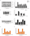
Inhibitory Role of Piperine in Activation of NADPH Oxidase-Derived Reactive Oxygen Species (ROS) during LCA-Induced IL-8 Expression in CRC Cells. (A) HCT-116 cells pretreated with N-acetyl-l-cysteine (NAC), diphenyleneiodonium chloride (DPI), or piperine for 1 h were incubated with 30 μM LCA for 10 min. Cells were then treated with 5 μg/mL of 5- and 6-carboxyl 2′,7′-dichlorodihydro-fluorescein diacetate (DCFDA) in the dark for 10 min. DCF fluorescence was imaged using a confocal laser scanning fluorescence microscope, and the statistically significant values of ROS production are presented. Scale bar: 100 μm. (B) Cells pretreated with NAC or DPI for 1 h were incubated with 30 μM LCA for 4 h, followed by mRNA extraction and RT-PCR to determine IL-8 expression. (C) HCT-116 cells were transiently transfected with 500 ng pGL2-IL-8 promoter–reporter construct. These transfected cells were pretreated with NAC or DPI for 1 h and incubated with 30 μΜ LCA for 4 h, and the luciferase activity was measured using a luminometer. Data represent the mean ± SEM from three experimental trials. * p < 0.05 versus control; # p < 0.05 versus LCA. (D) HCT-116 cells transfected with si-Con or si-p47phox were incubated with 30 μM LCA for 30 min; cell lysates were analyzed for p47phox level using western blotting. (E) HCT-116 cells was transfected with si-Con or si-p47phox were incubated with 30 μM LCA for 4 h, and IL-8 mRNA level was measured using RT-PCR. (F) The NADPH oxidase activity measured by piperine pretreatment and LCA treatment. Data represent the mean ± SEM from three experimental trials. * p < 0.05 versus control; # p < 0.05 versus LCA. (G) HCT-116 cells pretreated with AG, PP1, PP2, or piperine for 1 h were incubated with 30 μM LCA for 4 h, and cell lysates were analyzed for p47phox level using western blot analysis.
Figure 5
Figure 5
Effect of Inhibitors on Expression of EGFR, Src, ERK, and AKT in HCT-116 Cells. (A) HCT-116 cells pretreated with 10 μM AG1478 (AG), 10 μM PP2, 30 μM PD-98059 (PD), or 20 μM LY-294002 (LY) for 1 h were incubated with 30 μM LCA, and cell lysates were analyzed for the phosphorylation level of EGFR, Src, ERK1/2, and AKT using western blotting. (B) HCT-116 cells pretreated with PD for 1 h were incubated with 30 μM LCA, and cell lysates were analyzed for the phosphorylation level of c-Fos and c-Jun through western blot analysis. (C) HCT-116 cells pretreated with AG, PP2, PD, or LY for 1 h were treated with 30 μM LCA, and cell lysates were analyzed for the phosphorylation level of p65 using western blot analysis. HCT-116 cells were transiently transfected with AP-1 luciferase reporter construct (D) or NF-κB luciferase reporter construct (E). These transfected cells were pretreated with 2 μM SR-11302 (SR), 5 mM N-acetyl-l-cysteine (NAC), 30 μM PD, 20 μM LY, or 10 μM BAY-11-7082 (BAY) for 1 h and incubated with 30 μΜ LCA for 4 h, and the luciferase activity was measured using a luminometer. Data represent the mean ± SEM from three experimental trials. * p < 0.05 versus control; # p < 0.05 versus LCA.
Figure 6
Figure 6
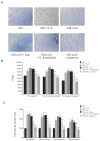
Effect of CM Obtained from the Piperine-Pretreated and LCA-Treated HCT-116 Cells on EA.hy926 for Tube Formation. EA.hy926 cells grown on a Matrigel-coated plate for 24 h were incubated with CM solutions. After 6 h, the cells were observed and counted using a Nokia microscope. (A) Representativeimages (10×) of the EA.hy926 tube formation. (B,C) Quantitative data of the EA. hy 926 tube formation. Data represent the mean ± SEM from three experimental trials. * p < 0.05 versus control; # p < 0.05 versus IL-8.
Figure 7
Figure 7
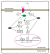
Schematic Representation of the Mechanism Underlying the Inhibitory Role of Piperine on LCA-Induced IL-8 Expression in CRC Cells and the Effect of CRC-Derived Angiogenesis in the Microenvironment. LCA induced IL-8 expression in human colorectal HCT-116 cells by increasing the transcriptional activity of AP-1 and NF-κB through the Src/EGFR-mediated ROS signaling pathways in HCT-116 cells. Piperine suppressed LCA-stimulated IL-8 expression by inhibiting the transcriptional activity of AP-1 and NF-κB and attenuating the Src/EGFR-mediated ROS-driven ERK1/2 and AKT signaling pathways. The CRC-derived IL-8 affects the endothelial EA.hy926 cell angiogenic activity in the microenvironment.

Similar articles

Cited by

References

    1. De Palma M., Biziato D., Petrova T.V. Microenvironmental regulation of tumour angiogenesis. Nat. Rev. Cancer. 2017;17:457–474. doi: 10.1038/nrc.2017.51. - DOI - PubMed
    1. Bielenberg D.R., Zetter B.R. The Contribution of Angiogenesis to the Process of Metastasis. Cancer J. 2015;21:267–273. doi: 10.1097/PPO.0000000000000138. - DOI - PMC - PubMed
    1. Ziyad S., Iruela-Arispe M.L. Molecular mechanisms of tumor angiogenesis. Genes Cancer. 2011;2:1085–1096. doi: 10.1177/1947601911432334. - DOI - PMC - PubMed
    1. Lugano R., Ramachandran M., Dimberg A. Tumor angiogenesis: Causes, consequences, challenges and opportunities. Cell. Mol. Life Sci. 2020;77:1745–1770. doi: 10.1007/s00018-019-03351-7. - DOI - PMC - PubMed
    1. Hwang Y.S., Jeong M., Park J.S., Kim M.H., Lee D.B., Shin B.A., Mukaida N., Ellis L.M., Kim H.R., Ahn B.W., et al. Interleukin-1beta stimulates IL-8 expression through MAP kinase and ROS signaling in human gastric carcinoma cells. Oncogene. 2004;23:6603–6611. doi: 10.1038/sj.onc.1207867. - DOI - PubMed