Review
. 2022 Feb 28;12(3):328.
doi: 10.3390/brainsci12030328.

A Breakdown of Immune Tolerance in the Cerebellum

Affiliations
Review

A Breakdown of Immune Tolerance in the Cerebellum

Christiane S Hampe et al. Brain Sci. .

Abstract

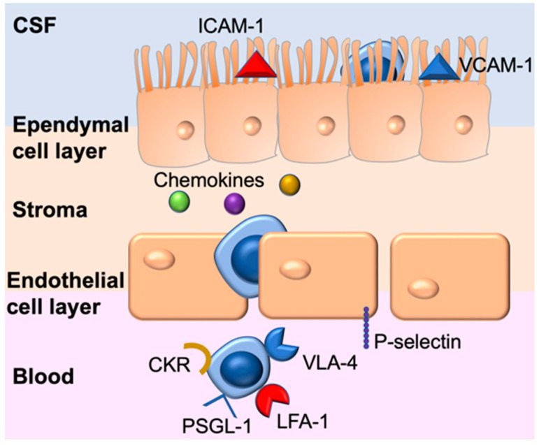
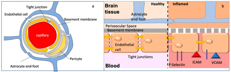
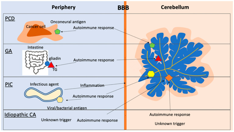
Cerebellar dysfunction can be associated with ataxia, dysarthria, dysmetria, nystagmus and cognitive deficits. While cerebellar dysfunction can be caused by vascular, traumatic, metabolic, genetic, inflammatory, infectious, and neoplastic events, the cerebellum is also a frequent target of autoimmune attacks. The underlying cause for this vulnerability is unclear, but it may be a result of region-specific differences in blood-brain barrier permeability, the high concentration of neurons in the cerebellum and the presence of autoantigens on Purkinje cells. An autoimmune response targeting the cerebellum-or any structure in the CNS-is typically accompanied by an influx of peripheral immune cells to the brain. Under healthy conditions, the brain is protected from the periphery by the blood-brain barrier, blood-CSF barrier, and blood-leptomeningeal barrier. Entry of immune cells to the brain for immune surveillance occurs only at the blood-CSF barrier and is strictly controlled. A breakdown in the barrier permeability allows peripheral immune cells uncontrolled access to the CNS. Often-particularly in infectious diseases-the autoimmune response develops because of molecular mimicry between the trigger and a host protein. In this review, we discuss the immune surveillance of the CNS in health and disease and also discuss specific examples of autoimmunity affecting the cerebellum.

Keywords: ataxia; autoimmunity; blood–CSF barrier; blood–brain barrier; cerebellum; immune surveillance.


Conflict of interest statement

The authors declare no conflict of interest.

Figures

Figure 1
Figure 1
Lymphocyte migration across the BCSFB. Lymphocytes expressing PSGL-1 interact with P-selectin constitutively expressed on the endothelial cell layer of the CP. In the stroma, lymphocytes expressing chemokine receptors (CKR) matching the chemokines present at the CP are attracted to migrate across the ependymal cell layer. In the CSF of the CP, lymphocytes can either attach via interactions between integrins LFA-1 and VLA-4 with the respective cell adhesion molecules ICAM-1 and VCAM-1, or remain in the CSF.
Figure 2
Figure 2
Blood–brain barrier. (a) Cellular components of the BBB. For details, see text. (b) Breakdown of BBB integrity. Inflammatory conditions induce the expression of P-selectin on the endothelial cells of the BBB, allowing PSGL-positive lymphocytes to tether. Tight junction proteins decrease in expression, and the basement membrane breaks down. Finally, the breakdown of the astrocyte end-feet allows access to the brain parenchyma.
Figure 3
Figure 3
Autoimmune cerebellar diseases. PCD: the onconeural antigen expressed on the cancer cells triggers an autoimmune response, which can recognize the same antigen in the cerebellum. PIC: The infectious agent evokes an immune response to an antigen that bears similarity to a neural antigen (mimicry). The infection also causes an inflammatory response, which compromises the BBB, allowing antibodies and immune cells access to the CNS. GA: Autoimmune response to the TG-gliadin complex results in TG-specific autoantibodies. TG autoantibodies can recognize brain-specific TGs. Idiopathic CA: the triggering event resulting in the autoimmune response is unclear. The trigger may occur in the periphery or in the CNS.

Similar articles

Cited by

References

    1. Bechmann I., Galea I., Perry V.H. What is the blood-brain barrier (not)? Trends Immunol. 2007;28:5–11. doi: 10.1016/j.it.2006.11.007. - DOI - PubMed
    1. Ousman S.S., Kubes P. Immune surveillance in the central nervous system. Nat. Neurosci. 2012;15:1096–1101. doi: 10.1038/nn.3161. - DOI - PMC - PubMed
    1. Engelhardt B., Vajkoczy P., Weller R.O. The movers and shapers in immune privilege of the CNS. Nat. Immunol. 2017;18:123–131. doi: 10.1038/ni.3666. - DOI - PubMed
    1. Garden G.A., Möller T. Microglia biology in health and disease. J. Neuroimmune Pharmacol. 2006;1:127–137. doi: 10.1007/s11481-006-9015-5. - DOI - PubMed
    1. Streit W.J., Mrak R.E., Griffin W.S.T. Microglia and neuroinflammation: A pathological perspective. J. Neuroinflamm. 2004;1:14. doi: 10.1186/1742-2094-1-14. - DOI - PMC - PubMed

LinkOut - more resources