

Tinnitus Perception in Light of a Parietal Operculo-Insular Involvement: A Review
- PMID: 35326290
- PMCID: PMC8946618
- DOI: 10.3390/brainsci12030334
Tinnitus Perception in Light of a Parietal Operculo-Insular Involvement: A Review
Abstract
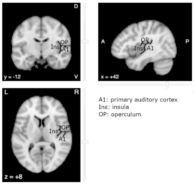
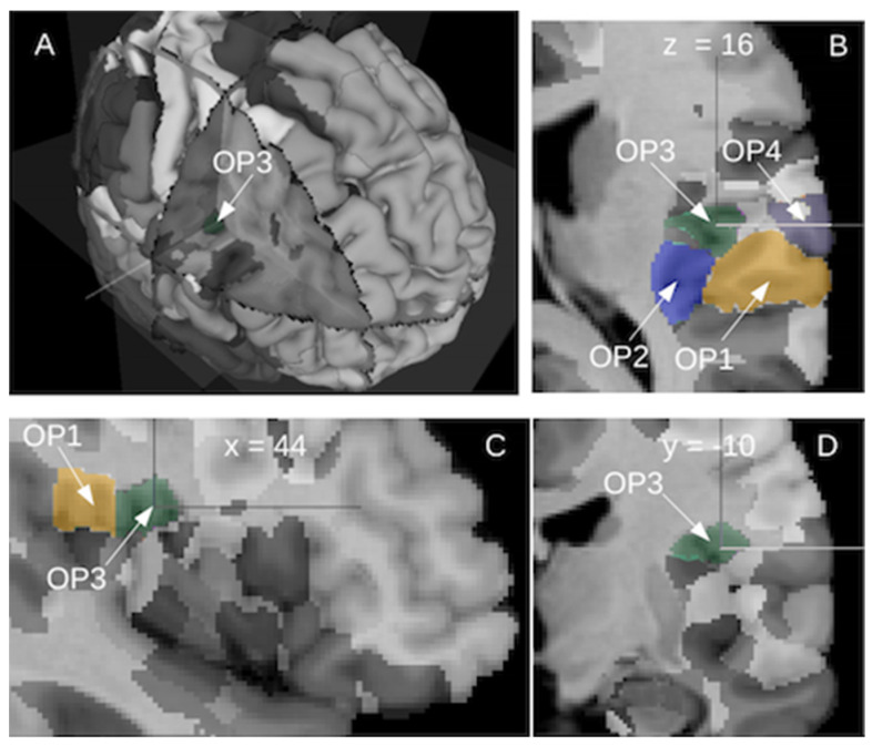
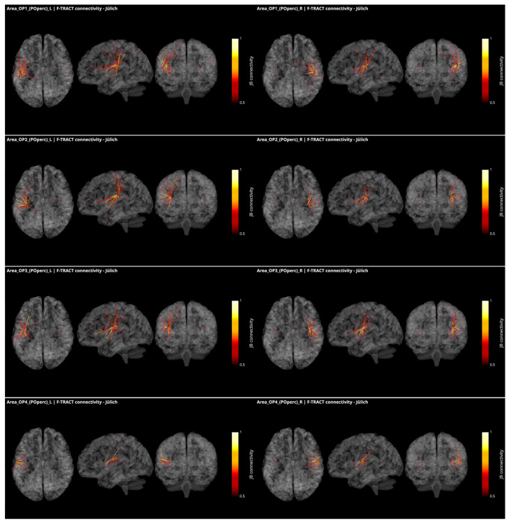
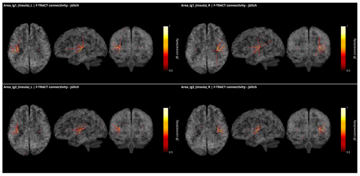
In tinnitus literature, researchers have increasingly been advocating for a clearer distinction between tinnitus perception and tinnitus-related distress. In non-bothersome tinnitus, the perception itself can be more specifically investigated: this has provided a body of evidence, based on resting-state and activation fMRI protocols, highlighting the involvement of regions outside the conventional auditory areas, such as the right parietal operculum. Here, we aim to conduct a review of available investigations of the human parietal operculo-insular subregions conducted at the microscopic, mesoscopic, and macroscopic scales arguing in favor of an auditory-somatosensory cross-talk. Both the previous literature and new results on functional connectivity derived from cortico-cortical evoked potentials show that these subregions present a dense tissue of interconnections and a strong connectivity with auditory and somatosensory areas in the healthy brain. Disrupted integration processes between these modalities may thus result in erroneous perceptions, such as tinnitus. More precisely, we highlight the role of a subregion of the right parietal operculum, known as OP3 according to the Jülich atlas, in the integration of auditory and somatosensory representation of the orofacial muscles in the healthy population. We further discuss how a dysfunction of these muscles could induce hyperactivity in the OP3. The evidence of direct electrical stimulation of this area eliciting auditory hallucinations further suggests its involvement in tinnitus perception. Finally, a small number of neuroimaging studies of therapeutic interventions for tinnitus provide additional evidence of right parietal operculum involvement.
Keywords: F-Tract; OP3; insula; integration area; neuroimaging; parietal operculum; tinnitus.
Conflict of interest statement
The funders had no role in the design of the study; in the collection, analyses, or interpretation of data; in the writing of the manuscript, or in the decision to publish the results.
Figures
Similar articles
-
Specific activation of operculum 3 (OP3) brain region during provoked tinnitus-related phantom auditory perceptions in humans.Brain Struct Funct. 2016 Mar;221(2):913-22. doi: 10.1007/s00429-014-0944-0. Epub 2014 Dec 12. Brain Struct Funct. 2016. PMID: 25503643
-
Functional Connectivity in Chronic Nonbothersome Tinnitus Following Acoustic Trauma: A Seed-Based Resting-State Functional Magnetic Resonance Imaging Study.Brain Connect. 2020 Aug;10(6):279-291. doi: 10.1089/brain.2019.0712. Epub 2020 Jul 17. Brain Connect. 2020. PMID: 32458713
-
Reduced sound-evoked and resting-state BOLD fMRI connectivity in tinnitus.Neuroimage Clin. 2018 Aug 31;20:637-649. doi: 10.1016/j.nicl.2018.08.029. eCollection 2018. Neuroimage Clin. 2018. PMID: 30202725 Free PMC article.
-
Tinnitus: Maladaptive auditory-somatosensory plasticity.Hear Res. 2016 Apr;334:20-9. doi: 10.1016/j.heares.2015.06.005. Epub 2015 Jun 12. Hear Res. 2016. PMID: 26074307 Free PMC article. Review.
-
Tinnitus Neural Mechanisms and Structural Changes in the Brain: The Contribution of Neuroimaging Research.Int Arch Otorhinolaryngol. 2015 Jul;19(3):259-65. doi: 10.1055/s-0035-1548671. Epub 2015 Mar 30. Int Arch Otorhinolaryngol. 2015. PMID: 26157502 Free PMC article. Review.
Cited by
-
Neural Plasticity in Tinnitus Mechanisms.Brain Sci. 2023 Nov 22;13(12):1615. doi: 10.3390/brainsci13121615. Brain Sci. 2023. PMID: 38137063 Free PMC article.
References
-
- Biswas R., Hall D.A. Prevalence, Incidence, and Risk Factors for Tinnitus. Curr. Top. Behav. Neurosci. 2021;51:3–28. - PubMed
-
- De Ridder D., Schlee W., Vanneste S., Londero A., Weisz N., Kleinjung T., Shekhawat G.S., Elgoyhen A.B., Song J.J., Andersson G., et al. Tinnitus and tinnitus disorder: Theoretical and operational definitions (an international multidisciplinary proposal) Prog. Brain Res. 2021;260:1–25. - PubMed
Publication types
Grants and funding
LinkOut - more resources
Full Text Sources
