. 2022 Mar 11;12(3):375.
doi: 10.3390/brainsci12030375.

Does the Heart Fall Asleep?-Diurnal Variations in Heart Rate Variability in Patients with Disorders of Consciousness

Affiliations

Does the Heart Fall Asleep?-Diurnal Variations in Heart Rate Variability in Patients with Disorders of Consciousness

Monika Angerer et al. Brain Sci. .

Abstract

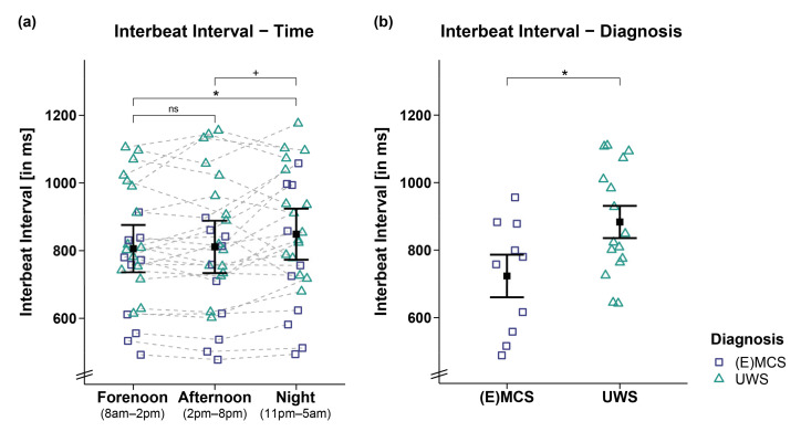
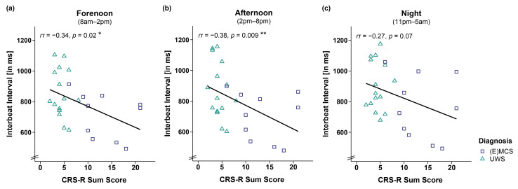
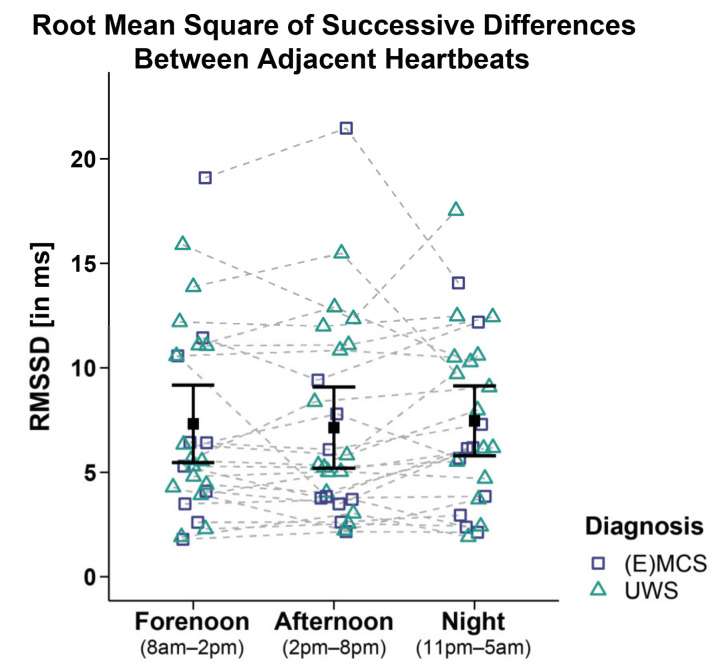
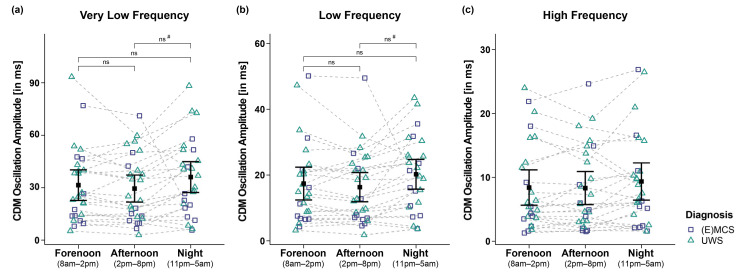
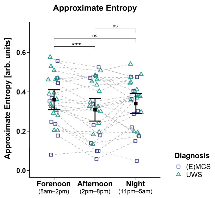
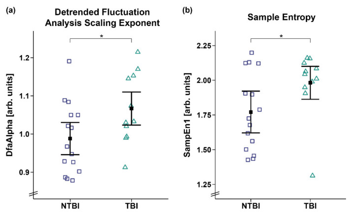
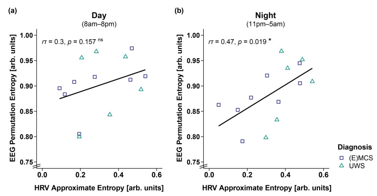
The current study investigated heart rate (HR) and heart rate variability (HRV) across day and night in patients with disorders of consciousness (DOC). We recorded 24-h electrocardiography in 26 patients with DOC (i.e., unresponsive wakefulness syndrome (UWS; n = 16) and (exit) minimally conscious state ((E)MCS; n = 10)). To examine diurnal variations, HR and HRV indices in the time, frequency, and entropy domains were computed for periods of clear day- (forenoon: 8 a.m.-2 p.m.; afternoon: 2 p.m.-8 p.m.) and nighttime (11 p.m.-5 a.m.). The results indicate that patients' interbeat intervals (IBIs) were larger during the night than during the day, indicating HR slowing. The patients in UWS showed larger IBIs compared to the patients in (E)MCS, and the patients with non-traumatic brain injury showed lower HRV entropy than the patients with traumatic brain injury. Additionally, higher HRV entropy was associated with higher EEG entropy during the night. Thus, cardiac activity varies with a diurnal pattern in patients with DOC and can differentiate between patients' diagnoses and etiologies. Moreover, the interaction of heart and brain appears to follow a diurnal rhythm. Thus, HR and HRV seem to mirror the integrity of brain functioning and, consequently, might serve as supplementary measures for improving the validity of assessments in patients with DOC.

Keywords: ECG; brain injury; disorders of consciousness; diurnal variation; heart rate; heart rate variability.


Conflict of interest statement

The authors declare no conflict of interest.

Figures

Figure 1
Figure 1
Interbeat interval (IBI) separately for time and diagnosis contrasts. (a) While patients’ IBIs were larger during the night as compared to the day (i.e., forenoon, afternoon), they did not differ between fore- and afternoon. (b) Patients in UWS showed larger IBIs than patients in (E)MCS. Error bars represent the mean and 95% confidence interval. * p < 0.05; + p ≤ 0.1; ns = not significant. Abbreviations: (E)MCS = (emergence from) minimally conscious state; UWS = unresponsive wakefulness syndrome; ms = milliseconds.
Figure 2
Figure 2
Correlation between interbeat interval (IBI) and CRS-R sum score separately for time. Larger IBIs were associated with lower CRS-R sum scores throughout the (a,b) day (i.e., forenoon, afternoon) and (c) night. Please note that the effect during the night is no longer ‘credible’ from a Bayesian point of view, and will not be interpreted. ** p < 0.01; * p < 0.05. Abbreviations: (E)MCS = (emergence from) minimally conscious state; UWS = unresponsive wakefulness syndrome; ms = milliseconds.
Figure 3
Figure 3
Root mean square of successive differences between adjacent heartbeats (RMSSD). Patients’ RMSSD did not differ between time ranges and diagnoses. Error bars represent the mean and 95% confidence interval. Abbreviations: (E)MCS = (emergence from) minimally conscious state; UWS = unresponsive wakefulness syndrome; ms = milliseconds.
Figure 4
Figure 4
Very low (VLF), low (LF), and high frequency (HF) oscillation amplitude of the IBI signal computed by complex demodulation (CDM). Analyses of the (a) VLF and (b) LF band revealed significant main effects for time (i.e., forenoon, afternoon, night). However, post hoc comparisons between times of day did not yield significance anymore after correction for multiple comparisons. (c) Patients’ HF also did not differ between times and diagnoses. Error bars represent the mean and 95% confidence interval. # = significant before correction for multiple comparisons; ns = not significant. Abbreviations: (E)MCS = (emergence from) minimally conscious state; UWS = unresponsive wakefulness syndrome; ms = milliseconds.
Figure 5
Figure 5
Approximate entropy (ApEn). Patients showed a higher ApEn during forenoon as compared to afternoon. No significant differences were evident between night and day (i.e., night vs. forenoon or afternoon). Error bars represent the mean and 95% confidence interval. *** p < 0.001; ns = not significant. Abbreviations: (E)MCS = (emergence from) minimally conscious state; UWS = unresponsive wakefulness syndrome; arb. units = arbitrary units.
Figure 6
Figure 6
Detrended fluctuation analysis scaling exponent (DfaAlpha) and sample entropy 1 (SampEn1). Patients with TBI showed a higher (a) DfaAlpha and (b) SampEn1 as compared to patients with NTBI. Error bars represent the mean and 95% confidence interval. * p < 0.05. Abbreviations: NTBI = non-traumatic brain injury; TBI = traumatic brain injury; arb. units = arbitrary units.
Figure 7
Figure 7
Correlation between EEG permutation entropy (PE) and HRV approximate entropy (ApEn) separately for day and night. (a) While EEG PE and HRV ApEn did not correlate during the day (i.e., 8 a.m.–8 p.m.), (b) a higher EEG PE was associated with a higher HRV ApEn during the night (i.e., 11 p.m.–5 a.m.). * p < 0.05; ns = not significant. Abbreviations: (E)MCS = (emergence from) minimally conscious state; UWS = unresponsive wakefulness syndrome; arb. units = arbitrary units.

Similar articles

Cited by

References

    1. Laureys S. The neural correlate of (un)awareness: Lessons from the vegetative state. Trends Cogn. Sci. 2005;9:556–559. doi: 10.1016/j.tics.2005.10.010. - DOI - PubMed
    1. Laureys S., Celesia G.G., Cohadon F., Lavrijsen J., Leon-Carrion J., Sannita W.G., Sazbon L., Schmutzhard E., von Wild K.R., Zeman A., et al. Unresponsive wakefulness syndrome: A new name for the vegetative state or apallic syndrome. BMC Med. 2010;8:1–4. doi: 10.1186/1741-7015-8-68. - DOI - PMC - PubMed
    1. Giacino J.T., Malone R. The vegetative and minimally conscious states. Handb. Clin. Neurol. 2008;90:99–111. doi: 10.1016/S0072-9752(07)01706-X. - DOI - PubMed
    1. Bruno M.A., Vanhaudenhuyse A., Thibaut A., Moonen G., Laureys S. From unresponsive wakefulness to minimally conscious PLUS and functional locked-in syndromes: Recent advances in our understanding of disorders of consciousness. J. Neurol. 2011;258:1373–1384. doi: 10.1007/s00415-011-6114-x. - DOI - PubMed
    1. Teasdale G., Jennett B. Assessment of coma and impaired consciousness. A practical scale. Lancet. 1974;2:81–84. doi: 10.1016/S0140-6736(74)91639-0. - DOI - PubMed

LinkOut - more resources