. 2022 Mar 10;11(6):949.
doi: 10.3390/cells11060949.

Vav1 Promotes B-Cell Lymphoma Development

Affiliations

Vav1 Promotes B-Cell Lymphoma Development

Batel Shalom et al. Cells. .

Abstract

Vav1 is normally and exclusively expressed in the hematopoietic system where it functions as a specific GDP/GTP nucleotide exchange factor (GEF), firmly regulated by tyrosine phosphorylation. Mutations and overexpression of Vav1 in hematopoietic malignancies, and in human cancers of various histologic origins, are well documented. To reveal whether overexpression of Vav1 in different tissues suffices for promoting the development of malignant lesions, we expressed Vav1 in transgenic mice by using the ubiquitous ROSA26 promoter (Rosa Vav1). We detected Vav1 expression in epithelial tissues of various organs including pancreas, liver, and lung. While carcinomas did not develop in these organs, surprisingly, we noticed the development of B-cell lymphomas. Rac1-GTP levels did not change in tissues from Rosa Vav1 mice expressing the transgenic Vav1, while ERK phosphorylation increased in the lymphomas, suggesting that signaling pathways are evoked. One of the growth factors analyzed by us as a suspect candidate to mediate paracrine stimulation in the lymphocytes was CSF-1, which was highly expressed in the epithelial compartment of Rosa Vav1 mice. The expression of its specific receptor, CSF-1R, was found to be highly expressed in the B-cell lymphomas. Taken together, our results suggest a potential cross-talk between epithelial cells expressing Vav1, that secrete CSF-1, and the lymphocytes that express CSF-1R, thus leading to the generation of B-cell lymphomas. Our findings provide a novel mechanism by which Vav1 contributes to tumor propagation.

Keywords: B-cell lymphoma; Rac-GTP; Rosa26; Vav1.


Conflict of interest statement

The authors have no competing interests to declare.

Figures

Figure 1
Figure 1
RosaVav1 transgenic mouse model (A) and expression pattern (B). (A) Schematic diagram of breeding strategy to generate RosaVav1 transgenic mice. Rosa26-rtTA mice were crossed with tetO-wtVav1 mice to generate Rosa26-rtTA/tetO-wtVav1 mice (Rosa Vav1 mice). To induce Vav1 expression, mice were treated with Dox one month after birth. Mice were sacrificed at different time points (6, 9, and 12 months after transgene induction). (B) Expression of transgenic Vav1 protein in the lungs of Rosa Vav1 mice. Western blotting using anti–GFP (indicative of the transgene humanVav1 expression) and anti-Actin Abs. (C) Expression of transgenic human Vav1 mRNA (GFP) and murine Vav1 (mVAV1) in the lungs of Rosa Vav1 mice. Quantitative real-time PCR analysis showing the mean mRNA expression of murine Vav1 (mVav1; left panel) and GFP (human transgenic Vav1; right panel) in lung tissues from Rosa Vav1 mice either treated (+Dox; n = 4) or non-treated (−Dox; n = 3), 12 months post transgene induction is depicted. SEM and significance between the treated (+Dox) and the non-treated (−Dox) analyzed by t-test are indicated. n.s. means non-significant.
Figure 2
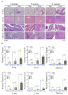
Figure 2
Appearance of Malignant lesions in Rosa Vav1 mice. (A) Representative images of hematoxylin and eosin (H&E) staining of lung, liver, pancreas, and spleen sections from Rosa Vav1 mice either treated (+Dox) or non-treated (−Dox), at indicated time points after transgene induction. Magnification at ×10. (B) Quantitative analysis of tumor numbers (upper panel) and percentage area (lower panel) in H&E-stained sections. The quantitation of area of tumors is detailed in the Material section. Number of mice used: non-treated (−Dox; 6, 9, 12 months n = 5); treated (+Dox; 6 months n = 5; 9 months n = 9; 12 months n = 12). SEM and significance between the treated (+Dox) and the non-treated (−Dox) at each time point analyzed by t-test are indicated.
Figure 3
Figure 3
Presence of B cells in the Lymphomas of Rosa Vav1 mice. Sections of lung, liver, pancreas, and spleen from Rosa Vav1 mice either treated (+Dox) or non-treated (−Dox) at indicated time points after transgene induction, were stained with H&E and anti-B220 antibodies. Representative pictures are shown. Magnification at ×10.
Figure 4
Figure 4
Expression of transgenic human Vav1(indicated by GFP) in the epithelial tissue at various organs of Rosa Vav1 mice. Sections of lung, liver, pancreas, and spleen from Rosa Vav1 mice either treated (+Dox) or non-treated (−Dox), Dox 12 months post transgene induction were stained with anti-GFP antibodies that identify the human Vav1 transgene (immunofluorescence; green) (A) or anti-Vav1 antibodies that identify murine and human Vav1 (B). Representative pictures are shown. Magnification at ×20.
Figure 5
Figure 5
Activation of ERK in Lymphomas from Rosa Vav1 mice. Analysis of Rac1 activation in the lung and ERK activation in the lung and liver of Rosa Vav1 mice either treated (+Dox) or non-treated (−Dox), 12 months post transgene induction was performed. (A) Protein lysates from _Rosa Vav1 transgenic mice were evaluated Rac1-GTP activation (upper panel; lung and liver tissues) and ERK phosphorylation (lower panels; lung and liver respectively) by using anti-Rac1-GTP, anti-Rac1, anti-pERK and anti-ERK Abs in western blotting. (B) The relative ratios of Rac1-GTP/Rac1 and pERK/ERK were calculated from the blots shown in A. The mean intensity of the western blots was quantified using ImageJ 1.49 V software. Numbers of mice used: non-treated (−Dox; n = 2); treated (+Dox; n = 5). SEM and significance between the treated (+Dox) and the non-treated (−Dox) analyzed by t-test are indicated. (C) Sections of lung (left) and liver (right) from indicated mice were stained with anti-pERK antibodies. Representative pictures are shown. Magnification at ×10.
Figure 6
Figure 6
Mechanism of generation of B-cell Lymphomas in Rosa Vav1 mice. (A) Sections of lung, liver, and pancreas from Rosa Vav1 mice either treated (+Dox) or non-treated (−Dox), 12 months post transgene induction stained with anti-Vav1, anti-CSF-1R and anti-CSF1 antibodies are depicted. Representative pictures are shown. Magnification at ×20. (B) The model proposed for generation of B-cell lymphomas from due to the aberrant expression of Rosa Vav1 in epithelial cells suggests that CSF-1 secretion from the epithelial compartment activates the CSF-1R on B-cells, leading eventually to the development of B-cell lymphoma.

References

    1. Katzav S., Martin-Zanca D., Barbacid M. Vav, a novel human oncogene derived from a locus ubiquitously expressed in hematopoietic cells. EMBO J. 1989;8:2283–2290. doi: 10.1002/j.1460-2075.1989.tb08354.x. - DOI - PMC - PubMed
    1. Katzav S. Flesh and blood: The story of Vav1, a gene that signals in hematopoietic cells but can be transforming in human malignancies. Cancer Lett. 2007;255:241–254. doi: 10.1016/j.canlet.2007.04.015. - DOI - PubMed
    1. Katzav S. Vav1: A hematopoietic signal transduction molecule involved in human malignancies. Int. J. Biochem. Cell Biol. 2009;41:1245–1248. doi: 10.1016/j.biocel.2008.11.006. - DOI - PubMed
    1. Tybulewicz V.L.J. Vav-family proteins in T-cell signalling. Curr. Opin. Immunol. 2005;17:267–274. doi: 10.1016/j.coi.2005.04.003. - DOI - PubMed
    1. Crespo P., Schuebel K.E., Ostrom A.A., Gutkind J.S., Bustelo X.R. Phosphotyrosine-dependent activation of Rac-1 GDP/GTP exchange by the vav proto-oncogene product. Nature. 1997;385:169–172. doi: 10.1038/385169a0. - DOI - PubMed

Publication types

Substances