

Fingolimod (FTY720), a Sphinogosine-1-Phosphate Receptor Agonist, Mitigates Choroidal Endothelial Proangiogenic Properties and Choroidal Neovascularization
- PMID: 35326420
- PMCID: PMC8946992
- DOI: 10.3390/cells11060969
Fingolimod (FTY720), a Sphinogosine-1-Phosphate Receptor Agonist, Mitigates Choroidal Endothelial Proangiogenic Properties and Choroidal Neovascularization
Abstract
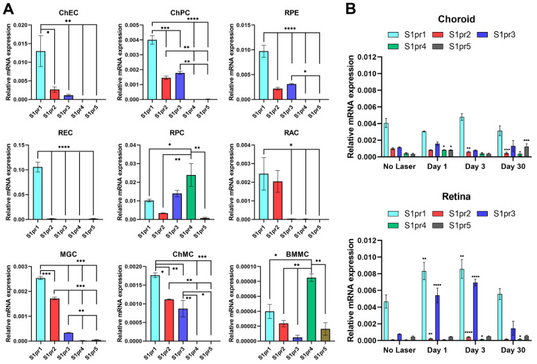
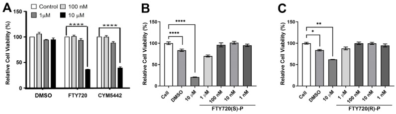
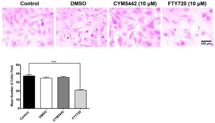
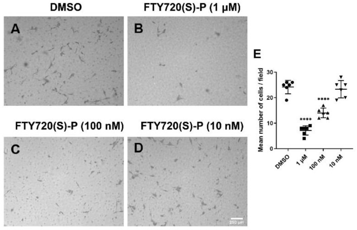
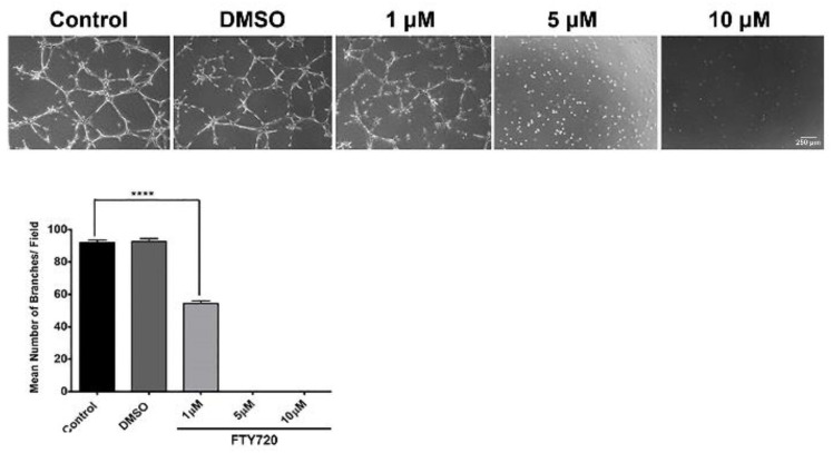
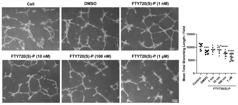
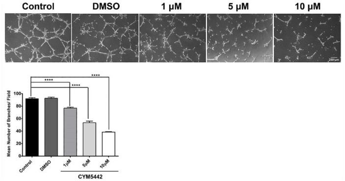
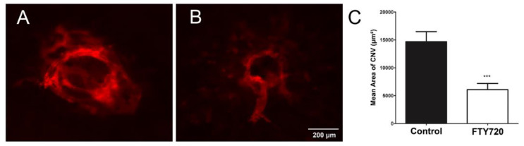
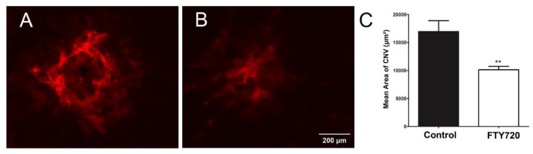
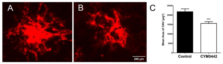
Neovascular or wet age-related macular degeneration (nAMD) causes vision loss due to inflammatory and vascular endothelial growth factor (VEGF)-driven neovascularization processes in the choroid. Due to the excess in VEGF levels associated with nAMD, anti-VEGF therapies are utilized for treatment. Unfortunately, not all patients have a sufficient response to such therapies, leaving few if any other treatment options for these patients. Sphingosine-1-phosphate (S1P) is a bioactive lipid mediator found in endothelial cells that participates in modulating barrier function, angiogenesis, and inflammation. S1P, through its receptor (S1PR1) in endothelial cells, prevents illegitimate sprouting angiogenesis during vascular development. In the present paper, we show that, in choroidal endothelial cells, S1PR1 is the most abundantly expressed S1P receptor and agonism of S1PR1-prevented choroidal endothelial cell capillary morphogenesis in culture. Given that nAMD pathogenesis draws from enhanced inflammation and angiogenesis as well as a loss of barrier function, we assessed the impact of S1PR agonism on choroidal neovascularization in vivo. Using laser photocoagulation rupture of Bruch's membrane to induce choroidal neovascularization, we show that S1PR non-selective (FTY720) and S1PR1 selective (CYM5442) agonists significantly inhibit choroidal neovascularization in this model. Thus, utilizing S1PR agonists to temper choroidal neovascularization presents an additional novel use for these agonists presently in clinical use for multiple sclerosis as well as other inflammatory diseases.
Keywords: age-related macular degeneration; choroid; inflammation; microglia; retinal pigment epithelium; retinal vasculature.
Conflict of interest statement
The authors state that the work presented here was not influenced by any commercial or financial relationships that could be considered as a potential conflict of interest.
Figures
Similar articles
-
ApoM-bound S1P acts via endothelial S1PR1 to suppress choroidal neovascularization and vascular leakage.Angiogenesis. 2025 Apr 23;28(2):24. doi: 10.1007/s10456-025-09975-7. Angiogenesis. 2025. PMID: 40266369 Free PMC article.
-
Artesunate mitigates choroidal neovascularization and scar formation.Exp Eye Res. 2023 Nov;236:109666. doi: 10.1016/j.exer.2023.109666. Epub 2023 Oct 4. Exp Eye Res. 2023. PMID: 37783334
-
Intrachoroidal neovascularization in transgenic mice overexpressing vascular endothelial growth factor in the retinal pigment epithelium.Am J Pathol. 2001 Mar;158(3):1161-72. doi: 10.1016/S0002-9440(10)64063-1. Am J Pathol. 2001. PMID: 11238064 Free PMC article.
-
Vascular endothelial growth factors in retinal and choroidal neovascular diseases.Ann Med. 2012 Feb;44(1):1-17. doi: 10.3109/07853890.2010.532150. Epub 2011 Feb 1. Ann Med. 2012. PMID: 21284527 Review.
-
Choroidal Neovascular Membranes in Retinal and Choroidal Tumors: Origins, Mechanisms, and Effects.Int J Mol Sci. 2023 Jan 5;24(2):1064. doi: 10.3390/ijms24021064. Int J Mol Sci. 2023. PMID: 36674579 Free PMC article. Review.
Cited by
-
Sphingosine-1-phosphate receptor 1/5 selective agonist alleviates ocular vascular pathologies.Sci Rep. 2024 Apr 27;14(1):9700. doi: 10.1038/s41598-024-60540-6. Sci Rep. 2024. PMID: 38678148 Free PMC article.
-
ApoM-bound S1P acts via endothelial S1PR1 to suppress choroidal neovascularization and vascular leakage.Angiogenesis. 2025 Apr 23;28(2):24. doi: 10.1007/s10456-025-09975-7. Angiogenesis. 2025. PMID: 40266369 Free PMC article.
-
Intracellular Signaling Pathways and Their Potential Targeting for Treatment of Ocular Posterior Segment Fibrosis.J Ophthalmic Vis Res. 2025 May 21;20:10.18502/jovr.v20.16966. doi: 10.18502/jovr.v20.16966. eCollection 2025. J Ophthalmic Vis Res. 2025. PMID: 40689128 Free PMC article. Review.
-
Macular edema after siponimod treatment for multiple sclerosis: a case report and literature review.BMC Neurol. 2023 Jul 31;23(1):286. doi: 10.1186/s12883-023-03333-0. BMC Neurol. 2023. PMID: 37525104 Free PMC article. Review.
-
Assessing the role of T cells in response to retinal injury to uncover new therapeutic targets for the treatment of retinal degeneration.J Neuroinflammation. 2023 Sep 9;20(1):206. doi: 10.1186/s12974-023-02867-x. J Neuroinflammation. 2023. PMID: 37689689 Free PMC article.
References
-
- Hernandez-Zimbron L.F., Zamora-Alvarado R., Ochoa-De la Paz L., Velez-Montoya R., Zenteno E., Gulias-Canizo R., Quiroz-Mercado H., Gonzalez-Salinas R. Age-related macular degeneration: New paradigms for treatment and management of amd. Oxidative Med. Cell. Longev. 2018;2018:8374647. doi: 10.1155/2018/8374647. - DOI - PMC - PubMed
-
- Fernández-Robredo P., Sancho A., Johnen S., Recalde S., Gama N., Thumann G., Groll J., García-Layana A. Current treatment limitations in age-related macular degeneration and future approaches based on cell therapy and tissue engineering. J. Ophthalmol. 2014;2014:510285. doi: 10.1155/2014/510285. - DOI - PMC - PubMed
Publication types
MeSH terms
Substances
Grants and funding
LinkOut - more resources
Full Text Sources
