. 2022 Mar 15;11(6):993.
doi: 10.3390/cells11060993.

Spermatozoal Mitochondrial Dynamics Markers and Other Functionality-Related Signaling Molecules Exert Circadian-like Response to Repeated Stress of Whole Organism

Affiliations

Spermatozoal Mitochondrial Dynamics Markers and Other Functionality-Related Signaling Molecules Exert Circadian-like Response to Repeated Stress of Whole Organism

Isidora M Starovlah et al. Cells. .

Abstract

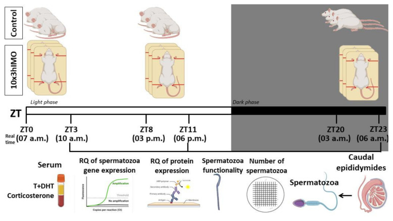
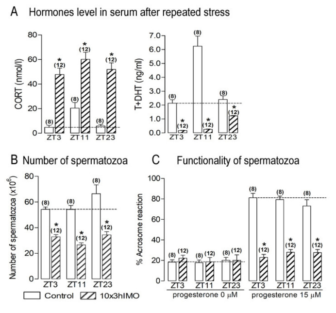
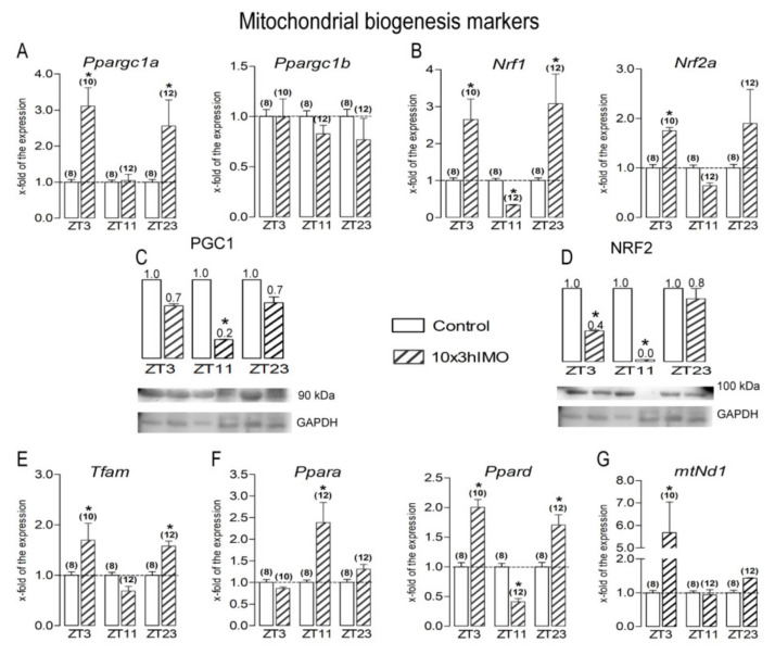
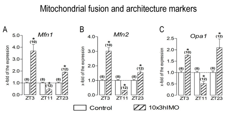
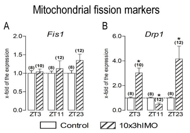
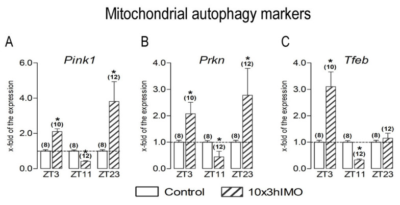
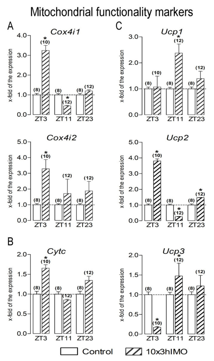
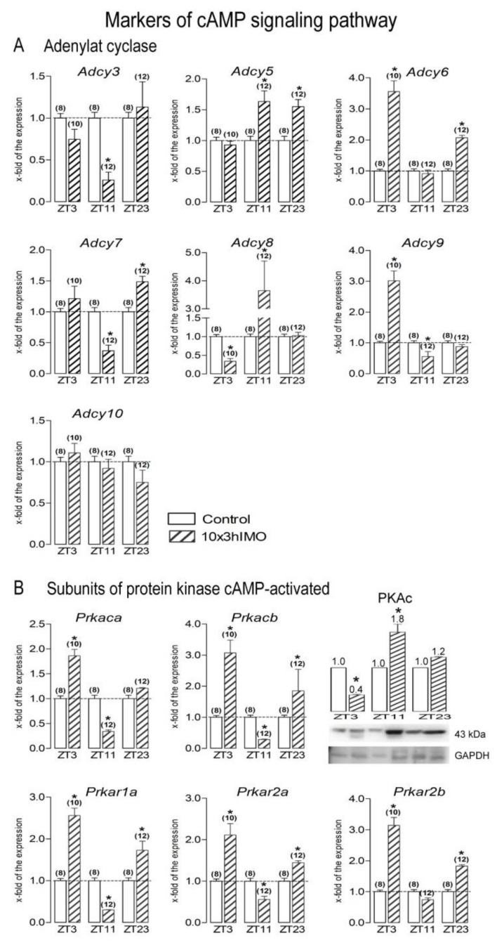
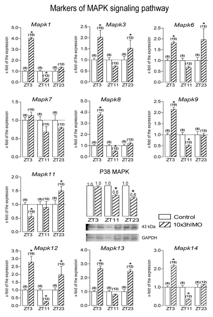
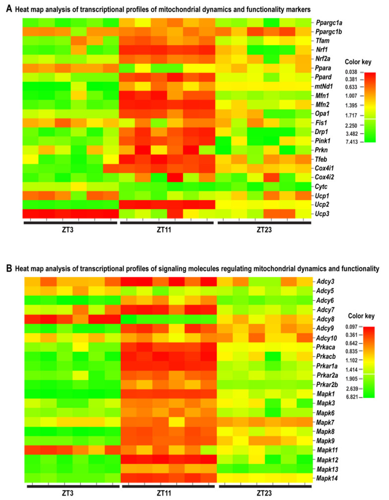
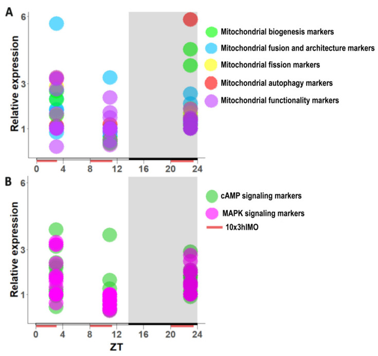
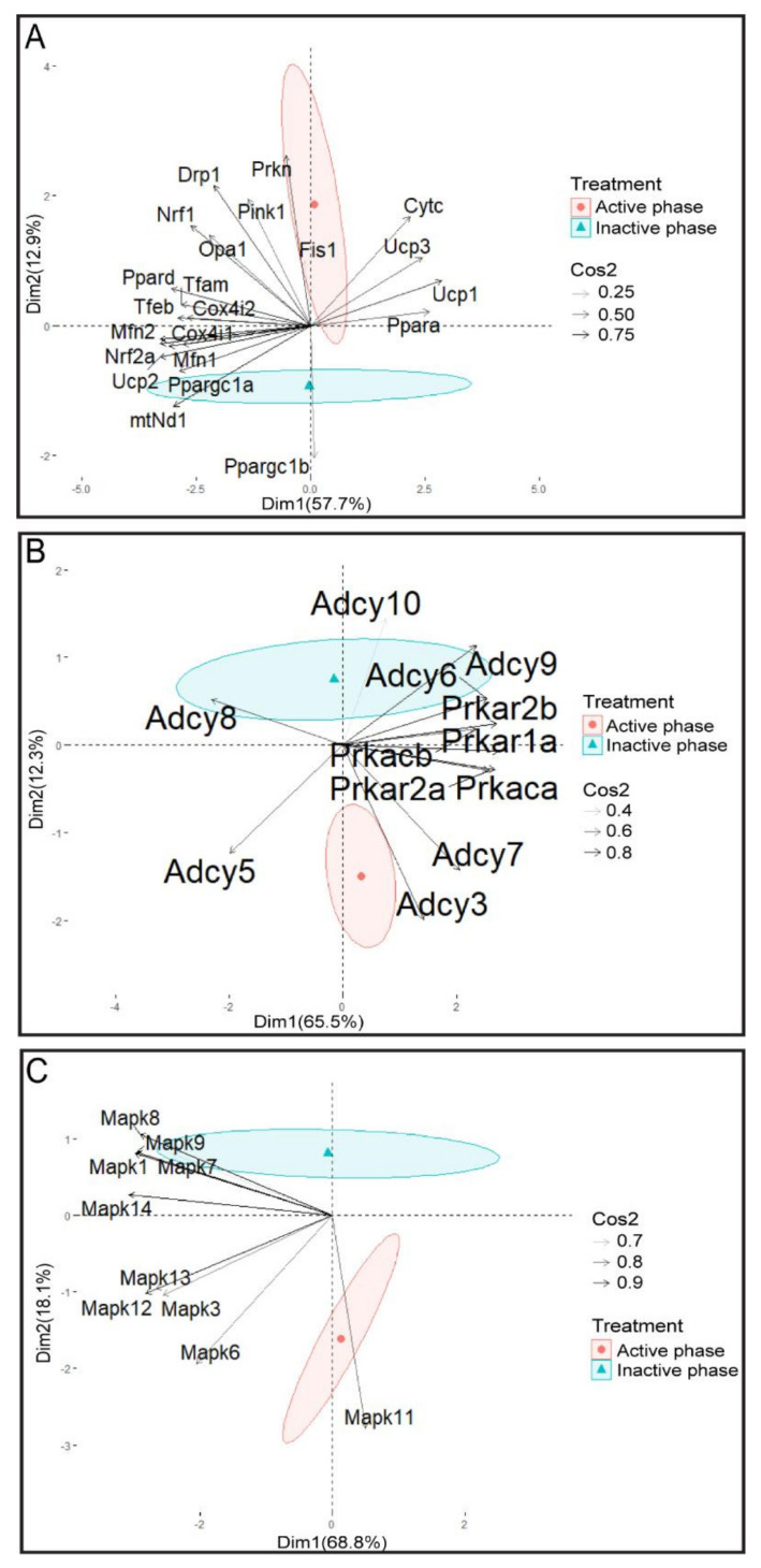
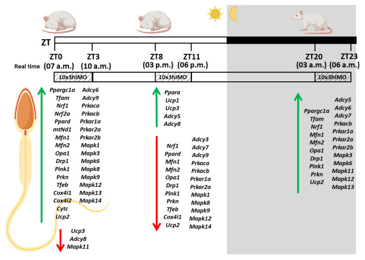
In the search for the possible role of the mitochondrial dynamics markers in spermatozoa adaptation, an in vivo approach was designed to mimic situations in which human populations are exposed to 3 h of repeated psychological stress (the most common stress in human society) at different time points during the day (24 h). The hormones (stress hormone corticosterone and testosterone), the number and the functionality of spermatozoa (response to acrosome-reaction-inducer progesterone), as well as the transcriptional profiles of 22 mitochondrial dynamics and function markers and 22 signaling molecules regulating both mitochondrial dynamics and spermatozoa number and functionality were followed at three time points (ZT3, ZT11, and ZT23). The results show that repeated stress significantly decreased the number and functionality of spermatozoa at all time points. In the same samples, the transcriptional profiles of 91% (20/22) of mitochondrial dynamics and functionality markers and 86% (19/22) of signaling molecules were disturbed after repeated stress. It is important to point out that similar molecular changes in transcriptional profiles were observed at ZT3 and ZT23, but the opposite was observed at ZT11, suggesting the circadian nature of the adaptive response. The results of PCA analysis show the significant separation of repeated stress effects during the inactive/light and active/dark phases of the day, suggesting the circadian timing of molecular adaptations.

Keywords: MAPK signaling markers; cAMP signaling markers; circadian; mitochondrial dynamics and functionality markers; repeated psychological stress response; spermatozoa number and functionality.


Conflict of interest statement

The authors declare no conflict of interest.

Figures

Figure 1
Figure 1
Experimental design of the in vivo experiment mimicking repeated stress applied at different time points during the day. Three-hour immobilization (IMO) stress was applied for 10 consecutive days (10×3hIMO) at different time points during the day (from ZT0 to ZT3, from ZT8 to ZT11, and from ZT20 to ZT23; ZT0 is the time when the light turned on). The relations of real time points with the ZT-time points: ZT3—3 h of stress started at 7 a.m. (ZT0; light on) and finished at 10 a.m.; ZT11—3 h of stress started at 3 p.m. (ZT8) and finished at 6 p.m.; ZT23—3 h of stress started next day at 3 a.m. (ZT20) and finished at 6 p.m. The levels of hormones, spermatozoa number and functionality (% acrosome reaction), as well as mitochondrial dynamics markers and related signaling molecules expressional profiles were followed. ZT—zeitgeber (time giver).
Figure 2
Figure 2
Repeated stress by immobilization (10×3hIMO) decreased androgen levels in circulation, together with functionality and number of spermatozoa, but increased the level of stress hormone corticosterone in different time points. Repeated psychophysical stress by immobilization (10×3hIMO) increased the circulating (A) stress hormone corticosterone level but decreased the level of androgens (testosterone + dihydrotestosterone, T+DHT). (B) The number of isolated spermatozoa from the caudal epididymides of undisturbed (control) rats and rats subjected to repeated immobilization stress, for 3 h for 10 consecutive days (10×3hIMO) in different periods during 24 h (from ZT0 to ZT3, from ZT8 to ZT11, and from ZT20 to ZT23; ZT0 was the time when the light turned on). (C) Spermatozoa functionality, presented as a % of acrosome-reacted spermatozoa, isolated from unstressed and repeatedly stressed (10×3hIMO) rats. After the capacitation, spermatozoa were stimulated with progesterone (PROG 15 µM) together with spermatozoa that were not treated with progesterone (PROG 0 µM). Acrosome-reacted spermatozoa were observed as the spermatozoa without the blue staining in the acrosome region, while blue staining in the acrosome region of the head of spermatozoa indicated intact acrosome. Data bars are mean ± SEM values of two independent in vivo experiments (n = number of rats). Statistical significance was set at level p < 0.05: * vs. control group of the same time point.
Figure 3
Figure 3
Different transcriptional profiles of mitochondrial biogenesis markers in spermatozoa of repeatedly stressed adult rats at different time points. Spermatozoa isolated from undisturbed and repeatedly stressed rats were used for RNA and protein isolation and further analysis of the transcriptional profile and protein expression profile of markers of mitochondrial biogenesis. The representative blots are shown as panels. Data from scanning densitometry were normalized to GAPDH (endogenous control). Values are shown as bars above the photos of blots, and numbers above the bars present fold of change. Data bars are mean ± SEM values of two independent in vivo experiments (n = number of rats). Statistical significance was set at level p < 0.05: * vs. control group of the same time point.
Figure 4
Figure 4
Different transcriptional profiles of mitochondrial fusion and architecture markers in spermatozoa of repeatedly stressed adult rats at different time points. Spermatozoa isolated from undisturbed and repeatedly stressed rats were used for RNA isolation and further analysis of the transcriptional profile of markers of mitochondrial fusion and architecture. Data bars are mean ± SEM values of two independent in vivo experiments (n = number of rats). Statistical significance was set at level p < 0.05: * vs. control group of the same time point.
Figure 5
Figure 5
Different transcriptional profiles of mitochondrial fission markers in spermatozoa of repeatedly stressed adult rats at different time points. Spermatozoa isolated from undisturbed and repeatedly stressed rats were used for RNA isolation and further analysis of the transcriptional profile of markers of mitochondrial fission. Data bars are mean ± SEM values of two independent in vivo experiments (n = number of rats). Statistical significance was set at level p < 0.05: * vs. control group of the same time point.
Figure 6
Figure 6
Different transcriptional profiles of mitochondrial autophagy markers in spermatozoa of repeatedly stressed adult rats at different time points. Spermatozoa isolated from undisturbed and repeatedly stressed rats were used for RNA isolation and further analysis of the transcriptional profile of markers of mitochondrial autophagy. Data bars are mean ± SEM values of two independent in vivo experiments (n = number of rats). Statistical significance was set at level p < 0.05: * vs. control group of the same time point.
Figure 7
Figure 7
Different transcriptional profiles of mitochondrial functionality markers in spermatozoa of repeatedly stressed adult rats at different time points. Spermatozoa isolated from undisturbed and repeatedly stressed rats were used for RNA isolation and further analysis of the transcriptional profile of markers of mitochondrial functionality. Data bars are mean ± SEM values of two independent in vivo experiments (n = number of rats). Statistical significance was set at level p < 0.05: * vs. control group of the same time point.
Figure 8
Figure 8
Different transcriptional profiles of markers of cAMP signaling regulating mitochondrial dynamics and functionality as well as spermatozoa number and functionality in spermatozoa of repeatedly stressed adult rats at different time points. Spermatozoa isolated from undisturbed and repeatedly stressed rats were used for RNA isolation and further analysis of the transcriptional profile of markers of cAMP signaling pathway. Data bars are mean ± SEM values of two independent in vivo experiments (n = number of rats). Statistical significance was set at level p < 0.05: * vs. control group of the same time point.
Figure 9
Figure 9
Different transcriptional profiles of markers of MAPK signaling regulating mitochondrial dynamics and functionality as well as spermatozoa number and functionality in spermatozoa of repeatedly stressed adult rats at different time points. Spermatozoa isolated from undisturbed and repeatedly stressed rats were used for RNA and protein isolation and further analysis of the transcriptional profile and protein expression profile of markers of MAPK signaling pathway. The representative blots are shown as panels. Data from scanning densitometry were normalized to GAPDH (endogenous control). Values are shown as bars above the photos of blots and numbers above the bars present fold of change. Data bars are mean ± SEM values of two independent in vivo experiments (n = number of rats). Statistical significance was set at level p < 0.05: * vs. control group of the same time point.
Figure 10
Figure 10
Heat map analysis of the transcriptional profile of mitochondrial dynamics and functionality markers (A) as well as signaling molecules regulating mitochondrial dynamics and functionality (B) in spermatozoa obtained from adult rats repeatedly stressed at different time points (ZT3, ZT11, and ZT23) during the day. Analysis showing different patterns of transcription at different time points in spermatozoa after repeated immobilization stress. Heat map analysis shows a relative fold of change in gene expression for the aforementioned markers at different time points (ZT3, ZT11, and ZT23), which are presented in colors from red to green, indicating low to high expression.
Figure 11
Figure 11
Pattern of transcripts from spermatozoa obtained from repeatedly stressed rats at different ZT time points (ZT3, ZT11, and ZT23) during the day (24 h). Data show transcriptional pattern of genes encoding the proteins are important for mitochondrial dynamics/functionality (A) as well as cAMP and MAPK signaling pathways (B). Points represent a deviation in the transcription of a particular gene at different ZT time points.
Figure 12
Figure 12
PCA of mitochondrial dynamics (A), cAMP signaling pathway (B), MAPK signaling pathway, (C) and gene expression on active/inactive phase; Dim1 and Dim2 represent the first two PCs and % of the retained variation. Cos2 estimates the qualitative representation of variables.
Figure 13
Figure 13
The transcriptional profile of mitochondrial dynamics and functionality markers as well as signaling molecules regulating mitochondrial dynamics and functionality in spermatozoa obtained from adult rats repeatedly stressed at different time points (ZT3, ZT11, and ZT23) during the day. Up arrow symbol represents increased transcription, while down arrow symbol represents decreased transcription.

Similar articles

References

    1. Markaki M., Tavernarakis N. Mitochondrial turnover and homeostasis in ageing and neurodegeneration. FEBS Lett. 2020;594:2370–2379. doi: 10.1002/1873-3468.13802. - DOI - PubMed
    1. Valenti D., Vacca R., Moro L., Atlante A. Mitochondria Can Cross Cell Boundaries: An Overview of the Biological Relevance, Pathophysiological Implications and Therapeutic Perspectives of Intercellular Mitochondrial Transfer. Int. J. Mol. Sci. 2021;22:8312. doi: 10.3390/ijms22158312. - DOI - PMC - PubMed
    1. Rosati A.J., Whitcomb B.W., Brandon N., Louis G.M.B., Mumford S.L., Schisterman E.F., Pilsner J.R. Sperm mitochondrial DNA biomarkers and couple fecundity. Hum. Reprod. 2020;35:2619–2625. doi: 10.1093/humrep/deaa191. - DOI - PMC - PubMed
    1. Ankel-Simons F., Cummins J.M. Misconceptions about mitochondria and mammalian fertilization: Implications for theories on human evolution. Proc. Natl. Acad. Sci. USA. 1996;93:13859–13863. doi: 10.1073/pnas.93.24.13859. - DOI - PMC - PubMed
    1. Hirata S., Hoshi K., Shoda T., Mabuchi T. Spermatozoon and mitochondrial DNA. Reprod. Med. Biol. 2002;1:41–47. doi: 10.1046/j.1445-5781.2002.00007.x. - DOI - PMC - PubMed

Publication types

LinkOut - more resources