. 2022 Mar 18;11(6):1038.
doi: 10.3390/cells11061038.

DHEA Protects Human Cholangiocytes and Hepatocytes against Apoptosis and Oxidative Stress

Affiliations

DHEA Protects Human Cholangiocytes and Hepatocytes against Apoptosis and Oxidative Stress

Ewa Kilanczyk et al. Cells. .

Abstract

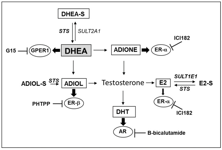
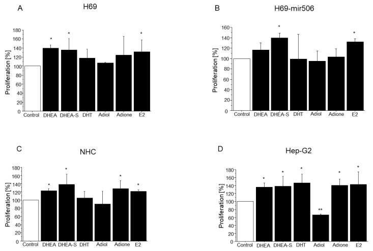
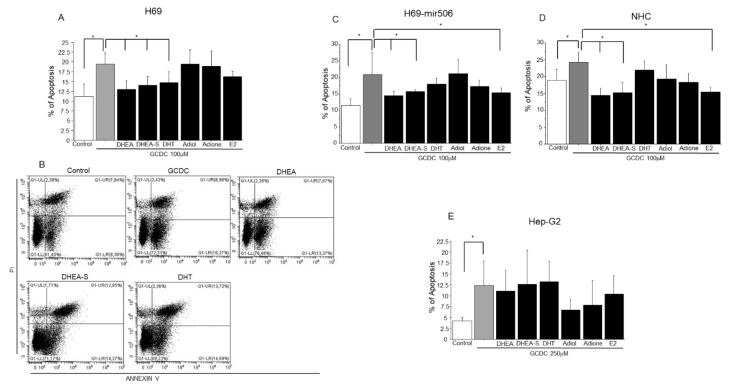
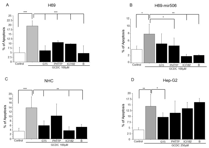
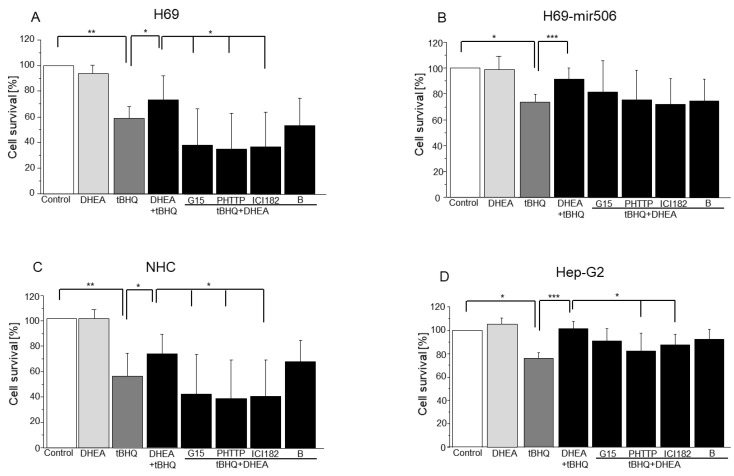
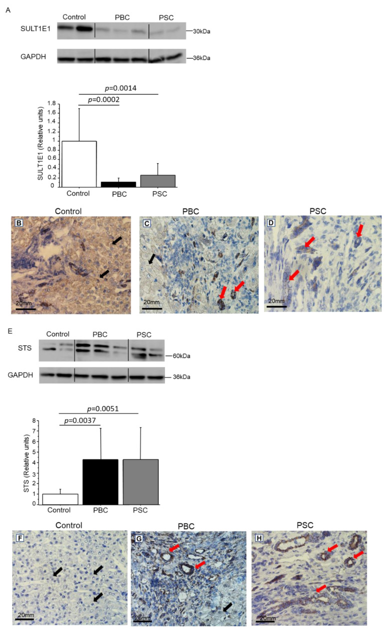
Primary biliary cholangitis (PBC) is a rare chronic cholestatic and immune-mediated liver disease of unknown aetiology that targets intrahepatic bile duct cells (cholangiocytes) and primarily affects postmenopausal women, when their estrogen levels sharply decrease. An impaired cholangiocyte response to estrogen characterizes the terminal stage of the disease, as this is when an inefficiency of cholangiocyte proliferation, in balancing the loss of intrahepatic bile ducts, is observed. Here, we report that the estrogen precursor dehydroepiandrosterone (DHEA) and its sulfate metabolites, DHEA-S and 17 β-estradiol, enhance the proliferation of cholangiocytes and hepatocytes in vitro. Flow cytometry analysis showed that DHEA and DHEA-S decreased glyco-chenodeoxycholic acid (GCDC)-driven apoptosis in cholangiocytes. Cell viability assay (MTT) indicated that ER-α, -β, and the G-protein-coupled estrogen receptor, are involved in the protection of DHEA against oxidative stress in cholangiocytes. Finally, immunoblot analysis showed an elevated level of steroid sulfatase and a reduced level of sulfotransferase 1E1 enzymes, involved in the desulfation/sulfation process of estrogens in cirrhotic PBC, and primary sclerosis cholangitis (PSC) liver tissues, another type of chronic cholestatic and immune-mediated liver disease. Taken together, these results suggest that DHEA can prevent the deleterious effects of certain potentially toxic bile acids and reactive oxygen species, delaying the onset of liver disease.

Keywords: DHEA; apoptosis; cholangiocytes.


Conflict of interest statement

The authors declare no conflict of interest.

Figures

Scheme 1
Scheme 1
Metabolic conversion of DHEA [24]. DHEA and its metabolites used in experiments are in square frames. AR—androgen receptor; B-bicalutamide—androgen receptor inhibitor; DHT—dihydrotestosterone; Adiol—androstenediol; Adione—androstenedione, ER—estrogen receptor; E2—17-β estradiol; G15—G-coupled estrogen receptor 1 (GPER1) inhibitor; ICI182—estrogen receptor α inhibitor; PHTPP—estrogen receptor β inhibitor; STS—steroid sulfatase; SULT2A1—sulfotransferase 2A1; SULT1E1—estrogen sulfotransferase.
Figure 1
Figure 1
The effect of DHEA and its metabolites on the proliferation of cholangiocytes (H69, H69-miR506, and NHC) and hepatocytes (Hep-G2). Different cells types i.e., cholangiocytes (AC) and hepatocytes (D) were incubated with DHEA and its metabolites (DHEA-S, DHT, adiol, adione and E2). Cells were harvested 48 h after treatment by scraping and were counted using CytoFLEX LX flow cytometer. Acquisition time was 60 s, and the sample flow rate was set at 60 μL/min. Results are presented as a mean ± SEM (n = 3); * p < 0.05; ** p < 0.01.
Figure 2
Figure 2
Effect of DHEA and its metabolites on the apoptosis induced by GCDC in cholangiocytes and hepatocytes. H69 (A,B), H69-mir506 (C), NHC (D) and Hep-G2 (E) cells were incubated with DHEA or its metabolites followed by GCDC. Twenty-four hours following treatment apoptosis was detected using an Annexin V/Propidium iodide (PI) kit. X-axis, Annexin V. Y-axis, PI fluorescence intensities. Representative figures show the population of viable (LL), early apoptotic (LR), late apoptotic (UR) and necrotic (UL) cells. Results are expressed as a percentage of early apoptotic cells from three separate experiments. * Statistically significant difference in comparison to cells treated with GCDC (* p < 0.05).
Figure 3
Figure 3
Involvement of estrogen and androgen receptors in the apoptosis induced by GCDC in cholangiocytes (H69, H69-miR506, NHC) and hepatocytes (Hep-G2). Cells (AD) were incubated with estrogen (GPER, ER-α, and ER-β) or androgen receptor inhibitors i.e., G15 (12 nM); ICI 182,780 (2 nM); PHTTP (10 nM), and bicalutamide (B) (10 nM), followed by GCDC treatment. After 24 h, cells were harvested by scraping and then incubated with Annexin V-FITC (1 ng/mL) and propidium iodide (5 ng/mL) for 30 min in the dark. They were analyzed by CytoFLEX LX flow cytometry, * p < 0.05; ** p < 0.01, *** p < 0.001.
Figure 4
Figure 4
The role of estrogen and androgen receptors in DHEA protection against mitochondrial oxidative stress induced by tBHQ. Cells (AD) were incubated with estrogen (GPER, ER-α, and ER-β) or androgen receptor inhibitors i.e., G15 (12 nM); ICI 182,780 (2 nM); PHTTP (10 nM), and bicalutamide (B) (10 nM), followed by DHEA and tBHQ co-treatment. To evaluate the viability of examined cells, MTT assays were conducted. Results are presented as mean ± SEM (n = 3); * p < 0.05, ** p < 0.01, *** p < 0.001.
Figure 5
Figure 5
The level and expression of SULT1E1 and STS in cirrhotic (PBC, PSC) and control liver tissues. Western blot analysis revealed lower levels of SULT1E1 (A) and higher levels of STS (E) in cirrhotic PBC or PSC tissues in comparison to controls. The levels of each protein were normalized to GAPDH as a loading control. Immunohistochemical analysis showed that in control and cirrhotic tissues SULT1E1 was primarily present in cholangiocytes within the bile ducts ((BD); red arrows) and in hepatocytes ((B,C); black arrows). Additionally, in cirrhotic tissues, STS was mainly present in cholangiocytes ((G,H); red arrows). In control tissues, this protein showed nuclear localization in hepatocytes (F). Scale bar—20 μm.

Similar articles

Cited by

References

    1. Alvaro D., Gigliozzi A., Attili A.F. Regulation and deregulation of cholangiocyte proliferation. J. Hepatol. 2000;33:333–340. doi: 10.1016/S0168-8278(00)80377-3. - DOI - PubMed
    1. Alvaro D., Invernizzi P., Onori P., Franchitto A., De Santis A., Crosignani A., Sferra R., Ginanni-Corradini S., Mancino M.G., Maggioni M., et al. Estrogen receptors in cholangiocytes and the progression of primary biliary cirrhosis. J. Hepatol. 2004;41:905–912. doi: 10.1016/j.jhep.2004.08.022. - DOI - PubMed
    1. Palmisano B.T., Zhu L., Stafford J.M. Role of Estrogens in the Regulation of Liver Lipid Metabolism. Adv. Exp. Med. Biol. 2017;1043:227–256. doi: 10.1007/978-3-319-70178-3_12. - DOI - PMC - PubMed
    1. Teng Y., Radde B.N., Litchfield L.M., Ivanova M.M., Prough R.A., Clark B.J., Doll M.A., Hein D.W., Klinge C.M. Dehydroepiandrosterone Activation of G-protein-coupled Estrogen Receptor Rapidly Stimulates MicroRNA-21 Transcription in Human Hepatocellular Carcinoma Cells. J. Biol. Chem. 2015;290:15799–15811. doi: 10.1074/jbc.M115.641167. - DOI - PMC - PubMed
    1. Van Erpecum K.J., Janssens A.R., Kreuning J., Ruiter D.J., Kroon H.M., Grond A.J. Generalized peliosis hepatis and cirrhosis after long-term use of oral contraceptives. Am. J. Gastroenterol. 1988;83:572–575. - PubMed

Publication types