

Emerging Roles of Airway Epithelial Cells in Idiopathic Pulmonary Fibrosis
- PMID: 35326501
- PMCID: PMC8947093
- DOI: 10.3390/cells11061050
Emerging Roles of Airway Epithelial Cells in Idiopathic Pulmonary Fibrosis
Abstract
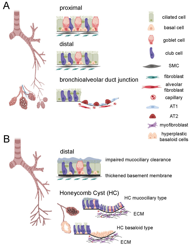
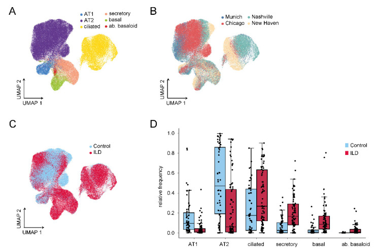
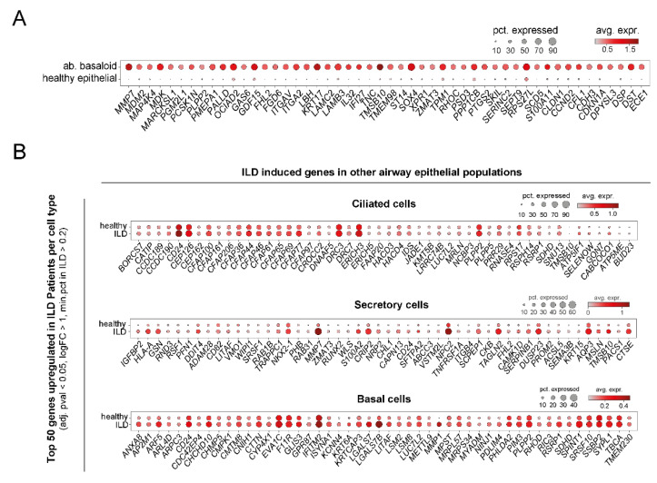
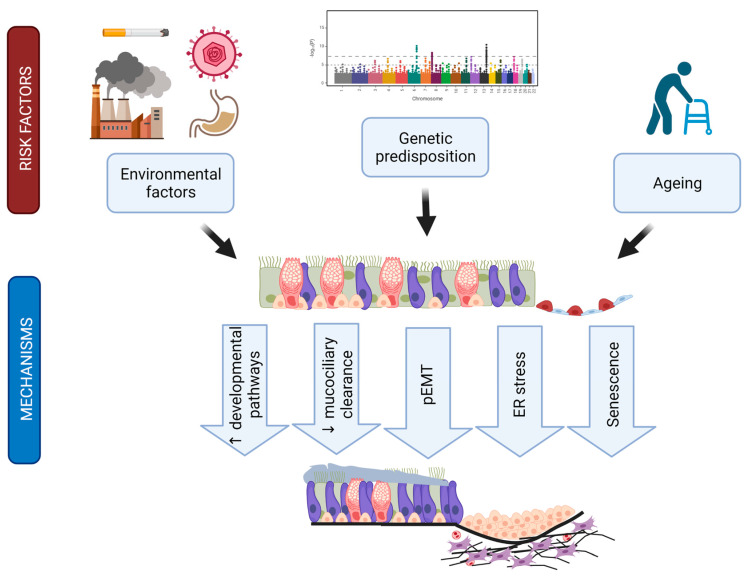
Idiopathic pulmonary fibrosis (IPF) is a fatal disease with incompletely understood aetiology and limited treatment options. Traditionally, IPF was believed to be mainly caused by repetitive injuries to the alveolar epithelium. Several recent lines of evidence, however, suggest that IPF equally involves an aberrant airway epithelial response, which contributes significantly to disease development and progression. In this review, based on recent clinical, high-resolution imaging, genetic, and single-cell RNA sequencing data, we summarize alterations in airway structure, function, and cell type composition in IPF. We furthermore give a comprehensive overview on the genetic and mechanistic evidence pointing towards an essential role of airway epithelial cells in IPF pathogenesis and describe potentially implicated aberrant epithelial signalling pathways and regulation mechanisms in this context. The collected evidence argues for the investigation of possible therapeutic avenues targeting these processes, which thus represent important future directions of research.
Keywords: IPF; MUC5B; airway epithelium; basal cells; bronchial epithelium; epithelial populations; lung fibrosis; single cell RNA sequencing.
Conflict of interest statement
The authors declare no conflict of interest. The funders had no role in the design of the study; in the collection, analyses, or interpretation of data; in the writing of the manuscript, or in the decision to publish the results.
Figures


Similar articles
-
The idiopathic pulmonary fibrosis honeycomb cyst contains a mucocilary pseudostratified epithelium.PLoS One. 2013;8(3):e58658. doi: 10.1371/journal.pone.0058658. Epub 2013 Mar 20. PLoS One. 2013. PMID: 23527003 Free PMC article.
-
Epithelial stem cells from human small bronchi offer a potential for therapy of idiopathic pulmonary fibrosis.EBioMedicine. 2025 Feb;112:105538. doi: 10.1016/j.ebiom.2024.105538. Epub 2025 Jan 2. EBioMedicine. 2025. PMID: 39753035 Free PMC article.
-
Single-cell RNA sequencing identifies diverse roles of epithelial cells in idiopathic pulmonary fibrosis.JCI Insight. 2016 Dec 8;1(20):e90558. doi: 10.1172/jci.insight.90558. JCI Insight. 2016. PMID: 27942595 Free PMC article.
-
Idiopathic Pulmonary Fibrosis: Pathogenesis and the Emerging Role of Long Non-Coding RNAs.Int J Mol Sci. 2020 Jan 14;21(2):524. doi: 10.3390/ijms21020524. Int J Mol Sci. 2020. PMID: 31947693 Free PMC article. Review.
-
Idiopathic Pulmonary Fibrosis Is a Genetic Disease Involving Mucus and the Peripheral Airways.Ann Am Thorac Soc. 2018 Nov;15(Suppl 3):S192-S197. doi: 10.1513/AnnalsATS.201802-144AW. Ann Am Thorac Soc. 2018. PMID: 30431344 Free PMC article. Review.
Cited by
-
Alveolar type 2 epithelial cell senescence and radiation-induced pulmonary fibrosis.Front Cell Dev Biol. 2022 Nov 2;10:999600. doi: 10.3389/fcell.2022.999600. eCollection 2022. Front Cell Dev Biol. 2022. PMID: 36407111 Free PMC article. Review.
-
Bronchial epithelial gene expression and interstitial lung abnormalities.Respir Res. 2023 Oct 10;24(1):245. doi: 10.1186/s12931-023-02536-w. Respir Res. 2023. PMID: 37817229 Free PMC article.
-
Precise identification of cell states altered in disease using healthy single-cell references.Nat Genet. 2023 Nov;55(11):1998-2008. doi: 10.1038/s41588-023-01523-7. Epub 2023 Oct 12. Nat Genet. 2023. PMID: 37828140 Free PMC article.
-
Dysregulated cross-talk between alveolar epithelial cells and stromal cells in idiopathic pulmonary fibrosis reduces epithelial regenerative capacity.Front Med (Lausanne). 2023 Aug 9;10:1182368. doi: 10.3389/fmed.2023.1182368. eCollection 2023. Front Med (Lausanne). 2023. PMID: 37621459 Free PMC article.
-
Single-cell RNA sequencing reveals special basal cells and fibroblasts in idiopathic pulmonary fibrosis.Sci Rep. 2024 Jul 9;14(1):15778. doi: 10.1038/s41598-024-66947-5. Sci Rep. 2024. PMID: 38982264 Free PMC article.
References
-
- King T.E., Jr., Bradford W.Z., Castro-Bernardini S., Fagan E.A., Glaspole I., Glassberg M.K., Gorina E., Hopkins P.M., Kardatzke D., Lancaster L., et al. A Phase 3 Trial of pirfenidone in patients with idiopathic pulmonary fibrosis. N. Engl. J. Med. 2014;370:2083–2092. doi: 10.1056/NEJMoa1402582. - DOI - PubMed
Publication types
MeSH terms
Grants and funding
LinkOut - more resources
Full Text Sources
Other Literature Sources
