. 2022 Mar 21;11(6):1055.
doi: 10.3390/cells11061055.

The Role of CD200-CD200 Receptor in Human Blood and Lymphatic Endothelial Cells in the Regulation of Skin Tissue Inflammation

Affiliations

The Role of CD200-CD200 Receptor in Human Blood and Lymphatic Endothelial Cells in the Regulation of Skin Tissue Inflammation

Dominic Rütsche et al. Cells. .

Abstract

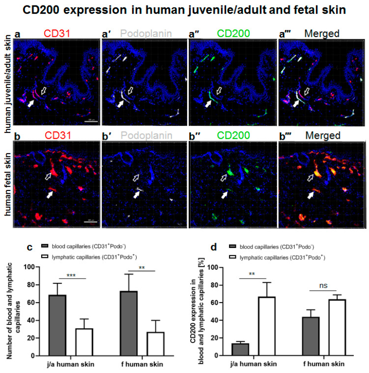
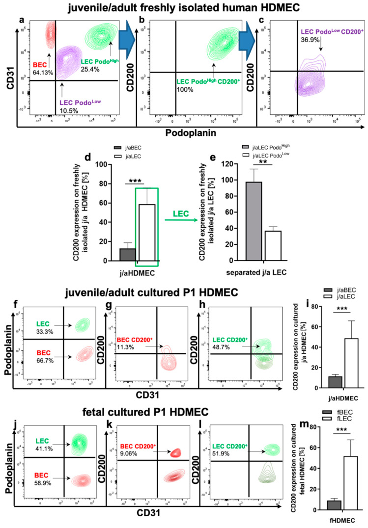
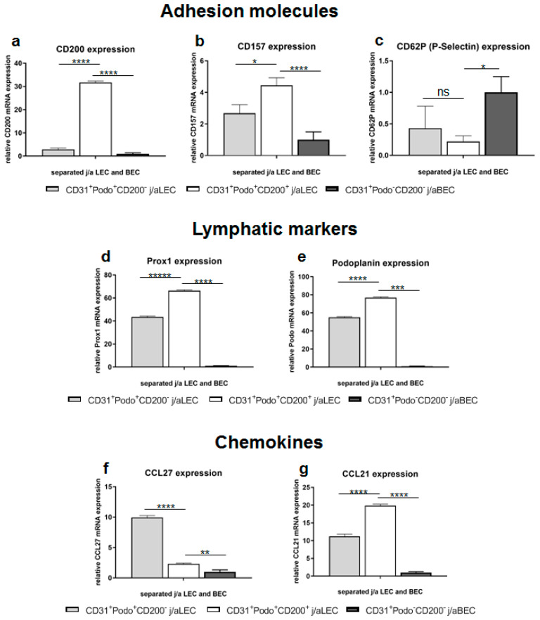
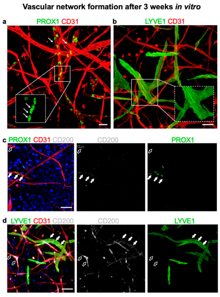
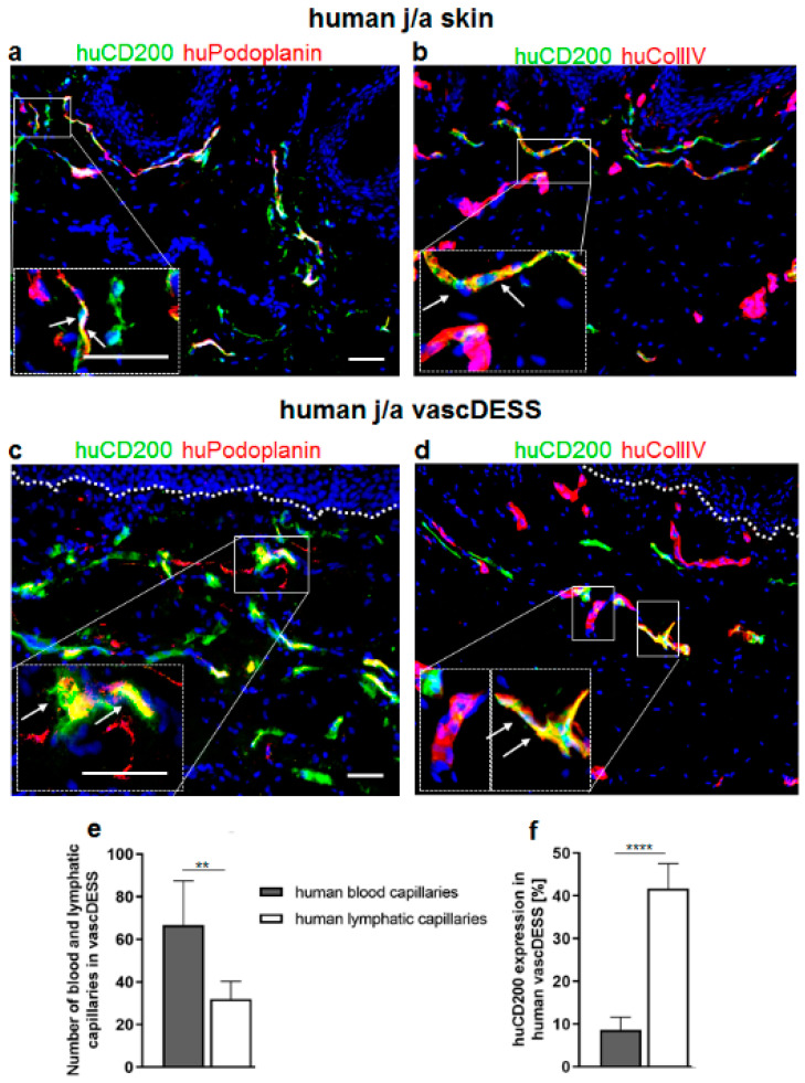
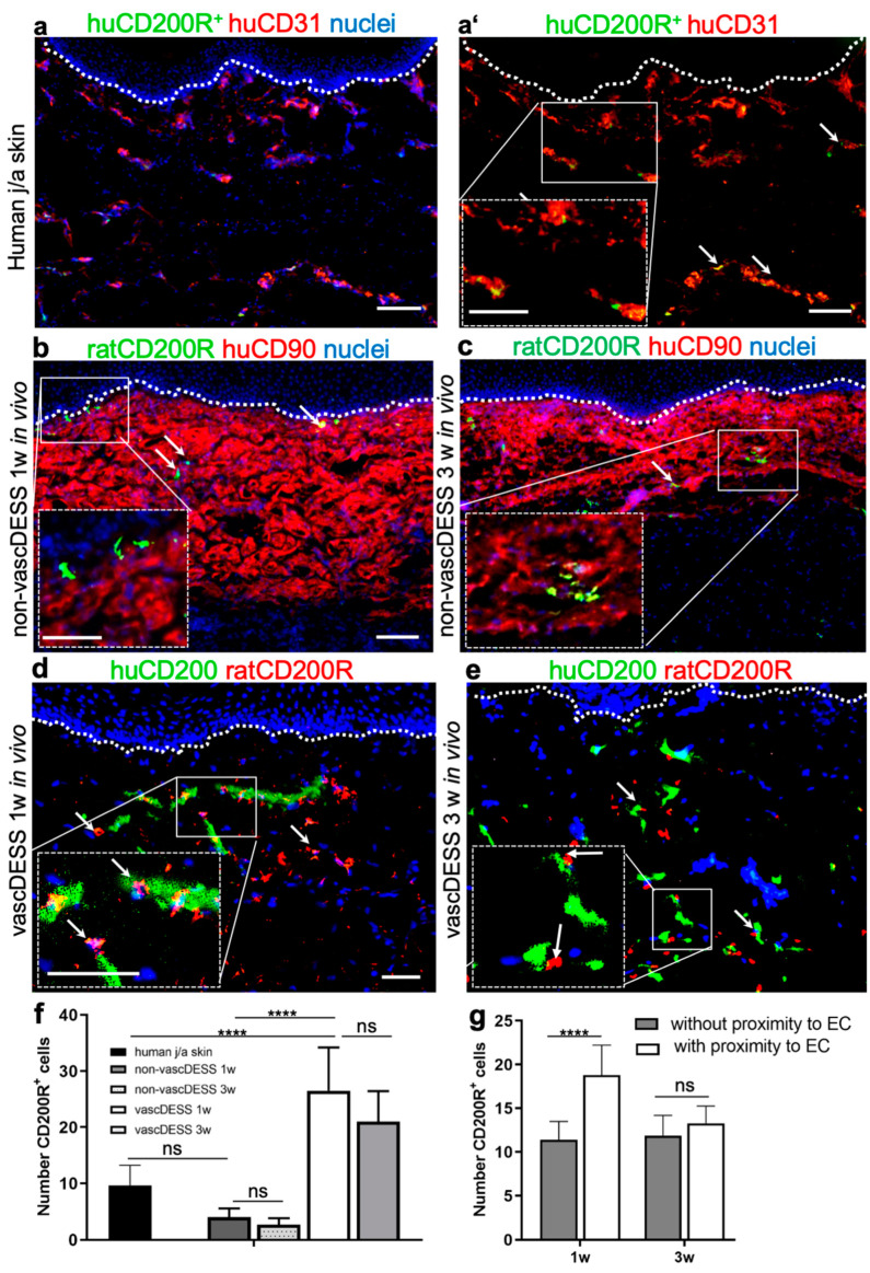
CD200 is a cell membrane glycoprotein that interacts with its structurally related receptor (CD200R) expressed on immune cells. We characterized CD200-CD200R interactions in human adult/juvenile (j/a) and fetal (f) skin and in in vivo prevascularized skin substitutes (vascDESS) prepared by co-culturing human dermal microvascular endothelial cells (HDMEC), containing both blood (BEC) and lymphatic (LEC) EC. We detected the highest expression of CD200 on lymphatic capillaries in j/a and f skin as well as in vascDESS in vivo, whereas it was only weakly expressed on blood capillaries. Notably, the highest CD200 levels were detected on LEC with enhanced Podoplanin expression, while reduced expression was observed on Podoplanin-low LEC. Further, qRT-PCR analysis revealed upregulated expression of some chemokines, including CC-chemokine ligand 21 (CCL21) in j/aCD200+ LEC, as compared to j/aCD200- LEC. The expression of CD200R was mainly detected on myeloid cells such as granulocytes, monocytes/macrophages, T cells in human peripheral blood, and human and rat skin. Functional immunoassays demonstrated specific binding of skin-derived CD200+ HDMEC to myeloid CD200R+ cells in vitro. Importantly, we confirmed enhanced CD200-CD200R interaction in vascDESS in vivo. We concluded that the CD200-CD200R axis plays a crucial role in regulating tissue inflammation during skin wound healing.

Keywords: CD200 (OX2); CD200 receptor; angiogenesis–immune cells/myeloid cells; blood capillaries; lymphatic capillaries; microvascular endothelial cells; regenerative medicine; skin bioengineering.


Conflict of interest statement

The authors declare no conflict of interest.

Figures

Figure 1
Figure 1
Evaluation of CD200 expression in normal human juvenile/adult and fetal skin. (aa’’’) Immunofluorescence triple staining of juvenile/adult (j/a) human skin with antibodies against CD31 (a) for human blood capillaries (red), podoplanin (a’) depicting human lymphatic endothelium (white), and CD200 (a’’) (green). (a’’’) represents a merged confocal Z stack. Whereas blood capillaries stain only CD31+ (empty arrows), lymphatic capillaries are CD31+Podo+ (white arrows). Note the presence of blood capillaries (CD31+Podo) which are mainly negative for CD200 (empty arrows), as well as the presence of lymphatic capillaries (CD31+Podo+) showing the expression of CD200 marker (white filled arrows). (bb’’’) Immunofluorescence triple staining of human fetal skin (f skin) against CD31 (b) delineating human blood capillaries (red), podoplanin (b’) depicting human lymphatic capillaries (white), and CD200 (b’’) (green). Note the presence of blood and lymphatic capillaries expressing CD200 (empty and filled white arrows, respectively). (b’’’) represents a merged overlay. (c) Quantification of blood and lymphatic capillaries in j/a and f skin. In the dermis of j/a, blood capillaries constitute approximately 68.8 ± 13%, whereas lymphatic endothelium represents 31.0 ± 10.6% of all vessels present in this skin type (*** p < 0.0001). In the f skin, blood endothelium comprises 73.0 ± 19% while lymphatic vessels constitute 27.0 ± 13% of all capillaries (** p < 0.001). (d) Quantification of CD200 expression on blood and lymphatic capillaries of j/a or f skin. Note the significantly higher expression of CD200 on j/a lymphatic capillaries as compared to the j/a blood capillaries (67.0 ± 16% vs. 14.0 ± 2.0%, respectively, *** p < 0.0001), whereas the expression of CD200 was similar in blood and lymphatic capillaries of human f skin (44.0 ± 8.0% vs. 64.0 ± 5.0%, respectively, p = ns), n = 5 independent j/a and fetal skin donors each. Cell nuclei are stained with Hoechst (blue). Scale bars 100 μm.
Figure 2
Figure 2
Expression of CD200 on freshly isolated as well as in vitro cultured juvenile/adult and fetal HDMEC. (a) Flow cytometry analysis of freshly isolated HDMEC derived from j/a human skin. CD31 and podoplanin were used to discriminate between blood endothelial cells (BEC, CD31+Podo) and lymphatic endothelial cells (LEC, CD31+Podo+). HDMEC obtained from freshly isolated j/a contained 60.13 ± 17.35% of j/aBEC. Note the presence of two distinct subpopulations of LEC expressing different levels of podoplanin, namely LEC PodoHigh, which comprise 26.8 ± 6.75%, as well as LEC PodoLow representing 11.5 ± 2.95% of all freshly isolated j/aHDMEC. (d,e) Flow cytometric analysis of CD200 expression in freshly isolated j/aBEC and PodoHigh and PodoLow LEC. Whereas only 12.91 ± 5.86% of freshly harvested j/aBEC stained positive for CD200, 58.73 ± 16.97% of all j/aLEC expressed this marker. Please note that LEC PodoHigh are almost entirely positive for CD200 (98.1 ± 15.6%) (b,e), while LEC PodoLow exhibit only a moderate expression of this marker (36.9 ± 5.1%) (p = 0.0041) (c,e). (fi) Flow cytometric analysis of cultured HDMEC at passage 1 (P1) derived from j/a skin. Cultured j/aHDMEC contains approximately 67.5 ± 18.21% of BEC as well as 31.8 ± 10.35% of LEC (f). Whereas 11.3 ± 2.0% of cultured j/aBEC exhibit CD200 expression (g,i), a significantly higher number of cultured j/aLEC was CD200-positive (48.7 ± 17.2, p < 0.0001) (h,i). (jm) Flow cytometric analysis of cultured fHDMEC at (P1) consisting of 58.4 ± 17.4% of fBEC and 41.6 ± 11.5 of fLEC. Further, 9.06 ± 2.0% of fBEC show the expression of CD200 (k,m), while 51.9 ± 15.7% of fLEC are positive for CD200 (l,m) (p < 0.0001). Two-way unpaired student t-test with ns = not significant (p > 0.05); ** for p-value 0.001 to 0.01 (very significant); and *** for p < 0.001 (extremely significant) was used for statistical analysis, n = 5 independent j/a and fetal skin donors each.
Figure 3
Figure 3
Proliferation rate of juvenille/adult (j/a) and fetal (f) CD200+/− LEC and BEC in vitro. (a) Graphical representation of proliferation rates of CD200+ and CD200 BEC and LEC isolated from j/a skin samples. Cells were plated at a concentration of 5 × 10³ cells per well (6-well plate), and their numbers were determined up to 11 days after plating by colorimetric cell counting assay. J/aCD200+ LEC (violet line) showed a higher proliferation rate in the first 5 days of the measurement as compared to j/aCD200 LEC (green line) and j/aBEC (red line). In turn, from day 7–11 during the assay, j/aBEC showed the highest cycling rate. At the same time, j/aCD200+ LEC showed lower proliferation rate as j/aCD200 LEC. Two-way ANOVA (analysis of variance) comparison of those two cell populations revealed following significance: d1/d5: p > 0.05 (ns); d3: p < 0.05; d7–d11: p < 0.001), whereas the comparison of j/aBEC vs. j/aCD200+ LEC demonstrated following values: d1/d3: 0.05 (ns); d5: p < 0.01; d7–d11: p < 0.001. (b) In days 1–5 of the assay, fCD200+ LEC (violet line) isolated from fetal skin revealed a slightly slower proliferation rate of as compared to fCD200 LEC (green line; d1/d5: p > 0.05 (ns); d3: p < 0.05), however it was higher as compared to fBEC (d1–d3: p > 0.05 (ns); d5 p < 0.01). Further, from day 5–11 the fCD200+ LEC population demonstrated the highest cycling rate as compared to fCD200 LEC (d5: p > 0.05 (ns); d7–d11: p < 0.001) and to fBEC (d5: p < 0.01; d7–d11: p < 0.001). Thus, fBEC showed a moderate proliferation rate with cycling values ranging between fCD200+ LEC and fCD200 LEC within days 5–11 of the assay. A representative experiment out of n = 6 (j/a) and n = 3 (f) is shown (n = different biological samples performed in triplicates). Two-way ANOVA with Bonferroni multiple comparisons test with ns = not significant (p > 0.05); * for p-value 0.01 to 0.05 (significant); *** for p < 0.001 (extremely significant) was used for statistical analysis (green means CD200+ LEC/CD200 LEC comparison; violet means CD200+ vs. BEC comparison). (c) Live-dead assay assessing viability at a 2-day interval in primary j/aHDMEC containing BEC/LEC. The data shows high cell viability even after confluence is reached in monolayer HDMEC culture. (d) Clonogenic assay of CD200+ and CD200 lymphatic endothelial cells (LEC). Quantification of the number of colonies revealed that CD31+Podo+CD200+ formed 32 ± 11.0 and CD31+Podo+CD200 37.7 ± 2.08 (p = 0.4351, ns) lymphatic endothelial cells formed similar numbers of colonies 14 days after cell seeding. n = 3 independent skin donors each. Experiments were conducted in triplicates. Scale bar 100 μm.
Figure 4
Figure 4
Gene expression analysis in distinct LEC and BEC fractions. HDMEC were harvested from j/a skin, cultured, separated at P0 into CD200/CD200+ LEC and CD200 BEC fractions and used directly for qRT-PCR. The relative gene expression levels were normalized to GAPDH housekeeping gene and displayed relative to CD200 BEC samples. (ac) Analysis of adhesion molecules confirmed 11-fold upregulation of CD200 mRNA in j/aCD200+ LEC (31.70 ± 0.67) as compared to j/aCD200 LEC (2.88 ± 0.6; p < 0.0001) and j/aCD200 BEC (1 ± 0.41; p < 0.0001) (a). Another adhesion molecule, CD157 was also enriched in j/aCD200+ LEC (4.44 ± 0.48) as compared to j/aCD200 LEC (2.68 ± 0.54; p = 0.0107) and j/aCD200 BEC (1 ± 0.5; p = 0.0003) (b). The expression of CD62P selectin mRNA was reduced in j/aCD200+ LEC (0.22 ± 0.09) in comparison to j/aCD200 LEC (0.43 ± 0.35; p = 0.5773, ns) and j/aCD200 BEC (1.0 ± 0.25; p = 0.0186) (c). (d,e) Assessment of mRNA expression of lymphatic markers. J/aCD200+ LEC showed upregulated expression of PROX1 (66.34 ± 0.74) and podoplanin (76.93 ± 0.77) as compared to j/aCD200 LEC (PROX1: 43.55 ± 0.78, podoplanin: 55.13 ± 0.76; both p < 0.0001) and j/aCD200 BEC (PROX1: 1 ± 0.41, podoplanin: 1 ± 0.09; both p < 0.0001). (f,g) Chemokines CCL27 and CCL21 showed various mRNA expressions, whereas CCL27 mRNA was downregulated in j/aCD200+ LEC (2.32 ± 0.11), CCL27 mRNA was upregulated in j/aCD200 LEC (9.96 ± 0.30; p < 0.0001) as compared to j/aCD200 BEC expression (1 ± 0.33; p = 0.0018). In contrast, CCL21 was significantly upregulated in j/aCD200+ LEC (19.86 ± 0.41), whereas CCL27 showed reduced mRNA levels in j/aCD200 LEC (11.17 ± 0.66; p < 0.0001). Each bar represents mean ± SD of n = 3 biological samples. Experimental groups were compared using one-way ANOVA with Turkey’s multiple comparisons test. Asterisks denote significance as follows: * p < 0.05; ** p < 0.01; *** p < 0.001; and **** p < 0.0001.
Figure 5
Figure 5
Cell adhesion assay of human immune cells to distinct endothelial cell (EC) populations. (ac) HUVEC (CD200) (a), j/aCD200 (b), and j/aCD200+ LEC (c) (all pre-labeled red) were co-cultured with separated human blood-derived granulocytes and T-cells (all pre-labeled green). (d) Cell adhesion assay shows a decreased population of pre-labeled human granulocytes and T-cells cells adhering to both CD200 cells: HUVEC (a) and j/aLEC (b) as compared to the high binding of both immune cell types to j/aCD200+ LEC (c) in vitro. (d) The quantification of the ratio of immune cells to EC confirmed that human blood-derived granulocytes and T-cells demonstrated specific adherence to j/aCD200+ LEC, whereas those immune cells did not show adherence to CD200 EC. Accordingly, the assessed adhesion ratio of CD200R+ granulocytes (9.77 ± 2.58) and T-cells (4.27 ± 1.59; p = 0.99 (ns)) to HUVEC was low and similar to the binding ratio to j/aCD200 LEC (11.11 ± 3.19 and 11.84 ± 0.93; p = 0.99 and p = 0.99 (ns), respectively). Further, the data showed that the adhesion rate of both immune cell types was significantly enhanced for j/aCD200+ LEC (91.27 ± 31.71 and 115.22 ± 29.69; p = 0.0005 and p < 0.0001, respectively). The results are expressed as mean ± SD from n = 6 biological samples of EC populations and n = 3 PBMC donors of granulocytes and T-cells. Asterisks denote significance as follows (unpaired t-test): *** p < 0.001. An unpaired t-test was used. Cell nuclei are stained with Hoechst (blue). Scale bars: 100 μm.
Figure 6
Figure 6
Expression of CD200 on in vitro cultured blood and lymphatic capillaries in 3D collagen. (a) Collagen type I-based hydrogel, and stained for endothelial-specific marker CD31 (red) and lymphatic lineage marker PROX1 (green) (n = 5). Whereas double-positive CD31+PROX1+ capillaries represent lymphatics, single positive CD31+PROX1 demonstrate blood capillaries. The lower inset shows a higher magnification of the single nuclear PROX1 staining in lymphatic capillaries (arrows). (b) The merged confocal immunofluorescence showing CD31+ blood capillaries (red) and lymphatic capillaries positive for CD31+ (red) and LYVE1 (green), which is a lymphatic-specific marker. Insets show a magnification of single-stained LYVE1 lymphatic capillaries. (c) Triple stained image of human capillary network co-stained for PROX1+ (green) CD200+ (white) CD31+ (red). Accordingly, PROX1+CD200+CD31+.triple positive capillaries represent lymphatics expressing CD200 marker (filled arrows), whereas double-positive PROX1CD200+CD31+ staining demonstrates blood capillaries positive for CD200 (empty arrows). (d) Triple co-staining for LYVE1+ (green) CD200+ (white) CD31+ (red). Whereas the majority of lymphatic capillaries (CD31+LYVE1+) were positive for CD200 (filled arrow), only a few blood capillaries (CD31+LYVE1) expressed CD200 (empty arrows). The results are representative from n = 3 biological samples of EC populations. Cell nuclei are stained with Hoechst (blue). Scale bars: (ad): 50 μm.
Figure 7
Figure 7
Quantification of CD200 expression in transplanted vascDESS. (a,b) Immunofluorescence co-staining of j/a human skin against huCD200 (green) and huPodoplanin (a) or collagen IV (b). Podoplanin depicts human lymphatic endothelium, while collagen IV is expressed exclusively around human blood capillaries. White arrows in magnification insets indicate double-positive microvascular structures. (c,d) Immunoflorescence co-staining of vascDESS against huCD200 (green) as well as huPodoplanin (red), visualizing the human lymphatic capillaries (c) and huCollagen IV (red) depicting the human blood endothelium (d). (e) Quantification revealed that vascDESS contains a significantly higher number of blood (66.59 ± 20.95) than lymphatic capillaries (32 ± 8.34; ** p = 0.009). Presented values are the mean (±SD) of total numbers of human CD31+ or podoplanin+ vessels counted per 10× high-power field, n = 6 biological samples in each group. White arrows in magnification insets indicate double-positive microvascular structures. (f) Quantification of CD200 expression on blood and lymphatic vessels in vascDESS. Please note that CD200 is expressed mainly on the lymphatic vessels (41.67 ± 5.89), while blood vessels exhibit only minor expression of that marker (8.63 ± 2.95; **** p < 0.0001). The results are expressed as mean ± SD from n = 3 biological donors from 3 independent animal transplantation (6 animals in total). Cell nuclei are stained with Hoechst (blue). Scale bars 50 μm.
Figure 8
Figure 8
Expression of CD200R in human j/a skin as well as in non-vascDESS and vascDESS. (a,a’) Immunofluorescence pictures of j/a skin stained against CD31 (red) and CD200R (green). Note that CD200R+ immune cells (9.65 ± 3.56) are closely associated with CD31+ capillaries. (b,c) The expression of CD200R in non-vascDESS at one week (4 ± 1.55, p = 0.1551 vs. normal skin) (b) and 3 weeks (2.67 ± 1.55, p = 0.0565 vs. normal skin) (c) after transplantation. Once again, CD200R-expressing cells were located in close proximity to the CD31+ capillaries. (d,e) The expression of CD200R in vascDESS one week (26.5 ± 7.71, p < 0.0001 vs. non-vascDESS 1w, **** p < 0.0001 vs. human skin) (d) and three weeks (21.31 ± 5.4, p < 0.0001 vs. non-vascDESS 1w, p < 0.0001 vs. human skin) (e) post-transplantation. Numerous CD200R+ cells were visible throughout the entire dermal layer and adhered mainly to the CD200+ endothelium. (f) Quantification of CD200R-expressing cells in the dermal compartments of j/a human skin as well as in non-vasc and vascDESS after one and three weeks in vivo. Please note the significantly higher number of CD200R+ cells in the dermis of vascDESS, as compared to the native skin or non-vascDESS. In contrast, human vascDESS containing human blood and lymphatic capillaries demonstrated enhanced density of rat CD200R-positive cells at one (26.5 ± 7.71, p < 0.0001 vs. non-vascDESS one week, **** p < 0.0001 vs. human skin) and three (21.31 ± 5.4, **** p < 0.0001 vs. non-vascDESS 1w, p < 0.0001 vs. human skin) weeks in vivo (d,e). White arrows in magnification insets indicate CD200–CD200R interactions. (g) In vivo, significantly more CD200+ endothelial cells were associated with rat CD200R+ immune cells 1 w post-transplantation compared to CD200R+ cells that were non-associated with CD200+ endothelial cells (**** p < 0.0001). This difference disappears after 3 w in vivo (ns, p > 0.05). The results are expressed as mean ± SD from n = 3 biological donors from 3 independent animal transplantation (6 animals in total). Cell nuclei are stained with Hoechst (blue). Scale bars 100 μm and 50 μm (magnification insets).

Similar articles

Cited by

References

    1. Biedermann T., Boettcher-Haberzeth S., Reichmann E. Tissue Engineering of Skin for Wound Coverage. Eur. J. Pediatr. Surg. 2013;23:375–382. doi: 10.1055/s-0033-1352529. - DOI - PubMed
    1. Biedermann T., Bottcher-Haberzeth S., Klar A.S., Widmer D.S., Pontiggia L., Weber A.D., Weber D.M., Schiestl C., Meuli M., Reichmann E. The Influence of Stromal Cells on the Pigmentation of Tissue-Engineered Dermo-Epidermal Skin Grafts. Tissue Eng. Part A. 2015;21:960–969. doi: 10.1089/ten.tea.2014.0327. - DOI - PMC - PubMed
    1. Bottcher-Haberzeth S., Biedermann T., Klar A.S., Widmer D.S., Neuhaus K., Schiestl C., Meuli M., Reichmann E. Characterization of pigmented dermo-epidermal skin substitutes in a long-term in vivo assay. Exp. Dermatol. 2015;24:16–21. doi: 10.1111/exd.12570. - DOI - PubMed
    1. Bottcher-Haberzeth S., Klar A.S., Biedermann T., Schiestl C., Meuli-Simmen C., Reichmann E., Meuli M. “Trooping the color”: Restoring the original donor skin color by addition of melanocytes to bioengineered skin analogs. Pediatr. Surg. Int. 2013;29:239–247. doi: 10.1007/s00383-012-3217-0. - DOI - PubMed
    1. Klar A.S., Biedermann T., Michalak K., Michalczyk T., Meuli-Simmen C., Scherberich A., Meuli M., Reichmann E. Human Adipose Mesenchymal Cells Inhibit Melanocyte Differentiation and the Pigmentation of Human Skin via Increased Expression of TGF-beta1. J. Invest. Dermatol. 2017;137:2560–2569. doi: 10.1016/j.jid.2017.06.027. - DOI - PubMed

Publication types

LinkOut - more resources