

Cytokine-Induced Senescence in the Tumor Microenvironment and Its Effects on Anti-Tumor Immune Responses
- PMID: 35326515
- PMCID: PMC8946098
- DOI: 10.3390/cancers14061364
Cytokine-Induced Senescence in the Tumor Microenvironment and Its Effects on Anti-Tumor Immune Responses
Abstract
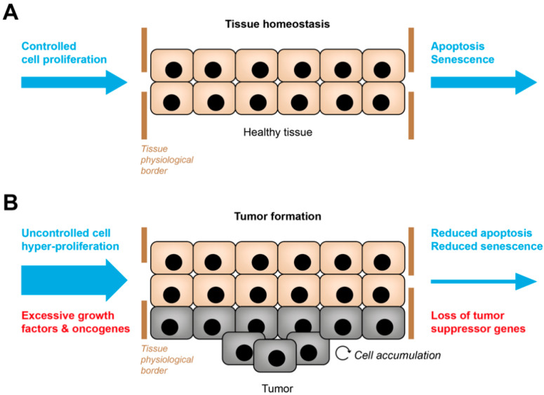
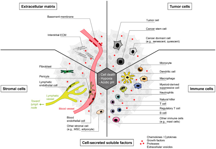
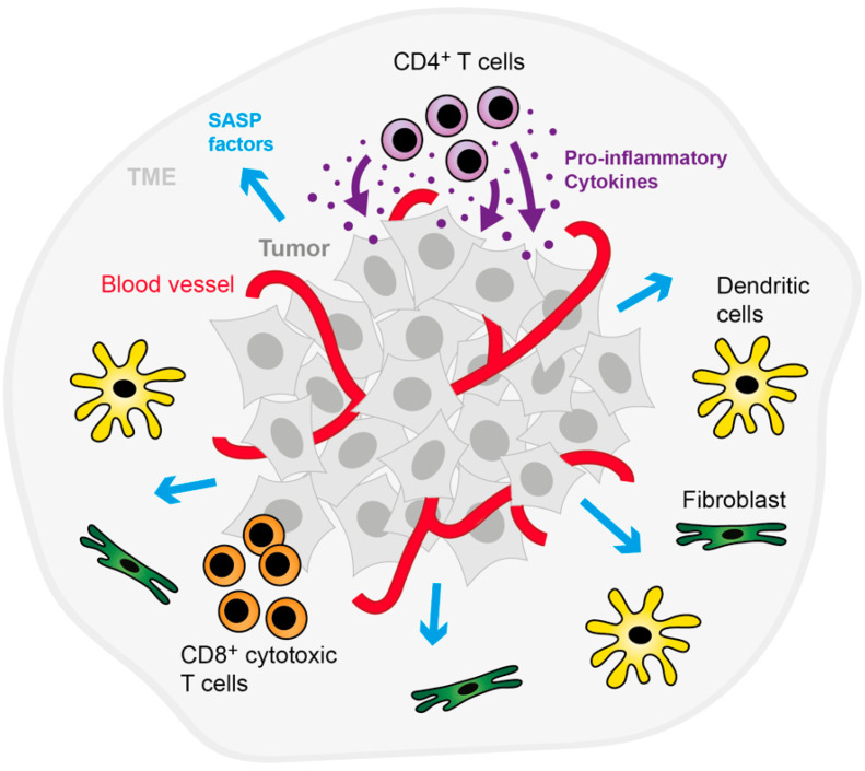
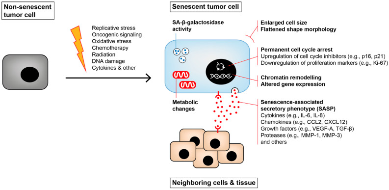
In contrast to surgical excision, chemotherapy or radiation therapy, immune checkpoint blockade therapies primarily influence cells in the tumor microenvironment, especially the tumor-associated lymphocytes and antigen-presenting cells. Besides complete remission of tumor lesions, in some patients, early tumor regression is followed by a consolidation phase where residing tumors remain dormant. Whereas the cytotoxic mechanisms of the regression phase (i.e., apoptosis, necrosis, necroptosis, and immune cell-mediated cell death) have been extensively described, the mechanisms underlying the dormant state are still a matter of debate. Here, we propose immune-mediated induction of senescence in cancers as one important player. Senescence can be achieved by tumor-associated antigen-specific T helper 1 cells, cytokines or antibodies targeting immune checkpoints. This concept differs from cytotoxic treatment, which often targets the genetic makeup of cancer cells. The immune system's ability to establish "defensive walls" around tumors also places the tumor microenvironment into the fight against cancer. Those "defensive walls" isolate the tumor cells instead of increasing the selective pressure. They also keep the tumor cells in a non-proliferating state, thereby correcting the derailed tissue homeostasis. In conclusion, strengthening the senescence surveillance of tumors by the immune cells of the microenvironment is a future goal to dampen this life-threatening disease.
Keywords: T cells; cell cycle regulation; cell death; chemoresistance; growth arrest; immunotherapy; inflammatory cytokines; senescence surveillance; tumor dormancy; tumor microenvironment.
Conflict of interest statement
The authors declare no conflict of interest.
Figures
Similar articles
-
Cytokine-induced senescence for cancer surveillance.Cancer Metastasis Rev. 2017 Jun;36(2):357-365. doi: 10.1007/s10555-017-9667-z. Cancer Metastasis Rev. 2017. PMID: 28391403 Review.
-
Therapy of lymphoma by immune checkpoint inhibitors: the role of T cells, NK cells and cytokine-induced tumor senescence.J Immunother Cancer. 2021 Jan;9(1):e001660. doi: 10.1136/jitc-2020-001660. J Immunother Cancer. 2021. PMID: 33441389 Free PMC article.
-
Changing T-cell enigma: cancer killing or cancer control?Cell Cycle. 2013 Oct 1;12(19):3146-53. doi: 10.4161/cc.26060. Epub 2013 Aug 26. Cell Cycle. 2013. PMID: 24013429 Free PMC article.
-
Immune checkpoint blockade therapy.J Allergy Clin Immunol. 2018 Nov;142(5):1403-1414. doi: 10.1016/j.jaci.2018.02.042. Epub 2018 Mar 27. J Allergy Clin Immunol. 2018. PMID: 29596939 Review.
-
Prospects for personalized combination immunotherapy for solid tumors based on adoptive cell therapies and immune checkpoint blockade therapies.Nihon Rinsho Meneki Gakkai Kaishi. 2017;40(1):68-77. doi: 10.2177/jsci.40.68. Nihon Rinsho Meneki Gakkai Kaishi. 2017. PMID: 28539557 Review.
Cited by
-
Counteracting Immunosuppression in the Tumor Microenvironment by Oncolytic Newcastle Disease Virus and Cellular Immunotherapy.Int J Mol Sci. 2022 Oct 27;23(21):13050. doi: 10.3390/ijms232113050. Int J Mol Sci. 2022. PMID: 36361831 Free PMC article. Review.
-
Comprehensive Analysis of Senescence Characteristics Defines a Novel Prognostic Signature to Guide Personalized Treatment for Clear Cell Renal Cell Carcinoma.Front Immunol. 2022 Jun 2;13:901671. doi: 10.3389/fimmu.2022.901671. eCollection 2022. Front Immunol. 2022. PMID: 35720278 Free PMC article.
-
IRF1 Mediates Growth Arrest and the Induction of a Secretory Phenotype in Alveolar Epithelial Cells in Response to Inflammatory Cytokines IFNγ/TNFα.Int J Mol Sci. 2024 Mar 19;25(6):3463. doi: 10.3390/ijms25063463. Int J Mol Sci. 2024. PMID: 38542436 Free PMC article.
-
Application of single-cell sequencing in the study of immune cell infiltration in inflammatory bowel disease and colorectal cancer.World J Gastrointest Oncol. 2025 Jun 15;17(6):107382. doi: 10.4251/wjgo.v17.i6.107382. World J Gastrointest Oncol. 2025. PMID: 40547160 Free PMC article. Review.
-
In Vitro Tumor Models on Chip and Integrated Microphysiological Analysis Platform (MAP) for Life Sciences and High-Throughput Drug Screening.Biosensors (Basel). 2023 Feb 6;13(2):231. doi: 10.3390/bios13020231. Biosensors (Basel). 2023. PMID: 36831997 Free PMC article. Review.
References
Publication types
Grants and funding
LinkOut - more resources
Full Text Sources
