

Hemodynamic Imaging in Cerebral Diffuse Glioma-Part A: Concept, Differential Diagnosis and Tumor Grading
- PMID: 35326580
- PMCID: PMC8946242
- DOI: 10.3390/cancers14061432
Hemodynamic Imaging in Cerebral Diffuse Glioma-Part A: Concept, Differential Diagnosis and Tumor Grading
Abstract
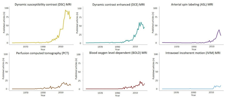
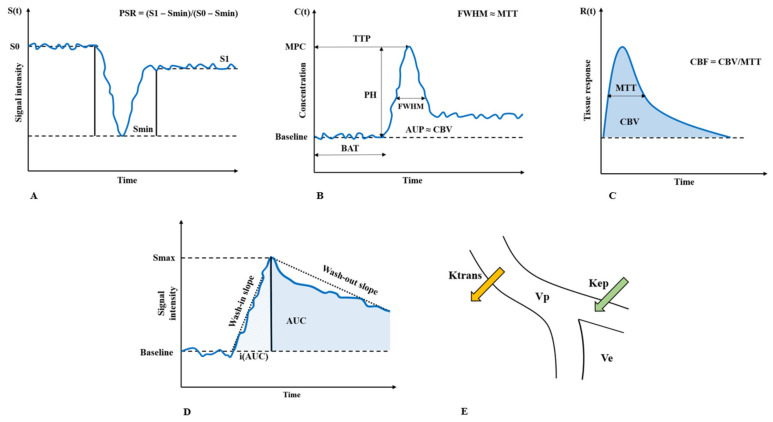
Diffuse gliomas are the most common primary malignant intracranial neoplasms. Aside from the challenges pertaining to their treatment-glioblastomas, in particular, have a dismal prognosis and are currently incurable-their pre-operative assessment using standard neuroimaging has several drawbacks, including broad differentials diagnosis, imprecise characterization of tumor subtype and definition of its infiltration in the surrounding brain parenchyma for accurate resection planning. As the pathophysiological alterations of tumor tissue are tightly linked to an aberrant vascularization, advanced hemodynamic imaging, in addition to other innovative approaches, has attracted considerable interest as a means to improve diffuse glioma characterization. In the present part A of our two-review series, the fundamental concepts, techniques and parameters of hemodynamic imaging are discussed in conjunction with their potential role in the differential diagnosis and grading of diffuse gliomas. In particular, recent evidence on dynamic susceptibility contrast, dynamic contrast-enhanced and arterial spin labeling magnetic resonance imaging are reviewed together with perfusion-computed tomography. While these techniques have provided encouraging results in terms of their sensitivity and specificity, the limitations deriving from a lack of standardized acquisition and processing have prevented their widespread clinical adoption, with current efforts aimed at overcoming the existing barriers.
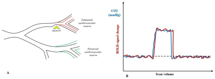
Keywords: MRI; cerebral glioma; cerebrovascular reactivity; glioblastoma; hemodynamic; perfusion MRI; perfusion computed tomography.
Conflict of interest statement
The authors declare no conflict of interest. The funders had no role in the design of the study; in the collection, analyses or interpretation of data; in the writing of the manuscript; or in the decision to publish the results.
Figures
Similar articles
-
Hemodynamic Imaging in Cerebral Diffuse Glioma-Part B: Molecular Correlates, Treatment Effect Monitoring, Prognosis, and Future Directions.Cancers (Basel). 2022 Mar 5;14(5):1342. doi: 10.3390/cancers14051342. Cancers (Basel). 2022. PMID: 35267650 Free PMC article. Review.
-
Quantification of blood flow in brain tumors: comparison of arterial spin labeling and dynamic susceptibility-weighted contrast-enhanced MR imaging.Radiology. 2003 Aug;228(2):523-32. doi: 10.1148/radiol.2282020409. Epub 2003 Jun 20. Radiology. 2003. PMID: 12819338
-
Imaging biomarkers guided anti-angiogenic therapy for malignant gliomas.Neuroimage Clin. 2018 Jul 5;20:51-60. doi: 10.1016/j.nicl.2018.07.001. eCollection 2018. Neuroimage Clin. 2018. PMID: 30069427 Free PMC article. Review.
-
Clinical Applications of Contrast-Enhanced Perfusion MRI Techniques in Gliomas: Recent Advances and Current Challenges.Contrast Media Mol Imaging. 2017 Mar 20;2017:7064120. doi: 10.1155/2017/7064120. eCollection 2017. Contrast Media Mol Imaging. 2017. PMID: 29097933 Free PMC article. Review.
-
Comparison of three different MR perfusion techniques and MR spectroscopy for multiparametric assessment in distinguishing recurrent high-grade gliomas from stable disease.Acad Radiol. 2013 Dec;20(12):1557-65. doi: 10.1016/j.acra.2013.09.003. Acad Radiol. 2013. PMID: 24200483
Cited by
-
Unlocking Bevacizumab's Potential: rCBVmax as a Predictive Biomarker for Enhanced Survival in Glioblastoma IDH-Wildtype Patients.Cancers (Basel). 2023 Dec 28;16(1):161. doi: 10.3390/cancers16010161. Cancers (Basel). 2023. PMID: 38201588 Free PMC article.
-
Cerebrovascular Reactivity Mapping in Brain Tumors Based on a Breath-Hold Task Using Arterial Spin Labeling.NMR Biomed. 2025 Mar;38(3):e5317. doi: 10.1002/nbm.5317. NMR Biomed. 2025. PMID: 39844376 Free PMC article.
-
Review of tracer kinetic models in evaluation of gliomas using dynamic contrast-enhanced imaging.Front Oncol. 2024 Jun 14;14:1380793. doi: 10.3389/fonc.2024.1380793. eCollection 2024. Front Oncol. 2024. PMID: 38947892 Free PMC article. Review.
-
Magnetic Resonance Imaging of Primary Adult Brain Tumors: State of the Art and Future Perspectives.Biomedicines. 2023 Jan 26;11(2):364. doi: 10.3390/biomedicines11020364. Biomedicines. 2023. PMID: 36830900 Free PMC article. Review.
-
Diffuse reflectance spectroscopy sensor to differentiate between glial tumor and healthy brain tissue: a proof-of-concept study.Biomed Opt Express. 2022 Nov 15;13(12):6470-6483. doi: 10.1364/BOE.474344. eCollection 2022 Dec 1. Biomed Opt Express. 2022. PMID: 36589562 Free PMC article.
References
-
- Weller M., van den Bent M., Preusser M., Le Rhun E., Tonn J.C., Minniti G., Bendszus M., Balana C., Chinot O., Dirven L., et al. EANO Guidelines on the Diagnosis and Treatment of Diffuse Gliomas of Adulthood. Nat. Rev. Clin. Oncol. 2020;18:170–186. doi: 10.1038/s41571-020-00447-z. - DOI - PMC - PubMed
-
- Jacobs D.I., Kumthekar P., Stell B.V., Grimm S.A., Rademaker A.W., Rice L., Chandler J.P., Muro K., Marymont M., Helenowski I.B., et al. Concordance of Patient and Caregiver Reports in Evaluating Quality of Life in Patients with Malignant Gliomas and an Assessment of Caregiver Burden. Neuro-Oncol. Pract. 2014;1:47–54. doi: 10.1093/nop/npu004. - DOI - PMC - PubMed
Publication types
Grants and funding
LinkOut - more resources
Full Text Sources
Research Materials
