Review
. 2022 Mar 12;14(6):1462.
doi: 10.3390/cancers14061462.

Chemotherapy Resistance: Role of Mitochondrial and Autophagic Components

Affiliations
Review

Chemotherapy Resistance: Role of Mitochondrial and Autophagic Components

Entaz Bahar et al. Cancers (Basel). .

Abstract

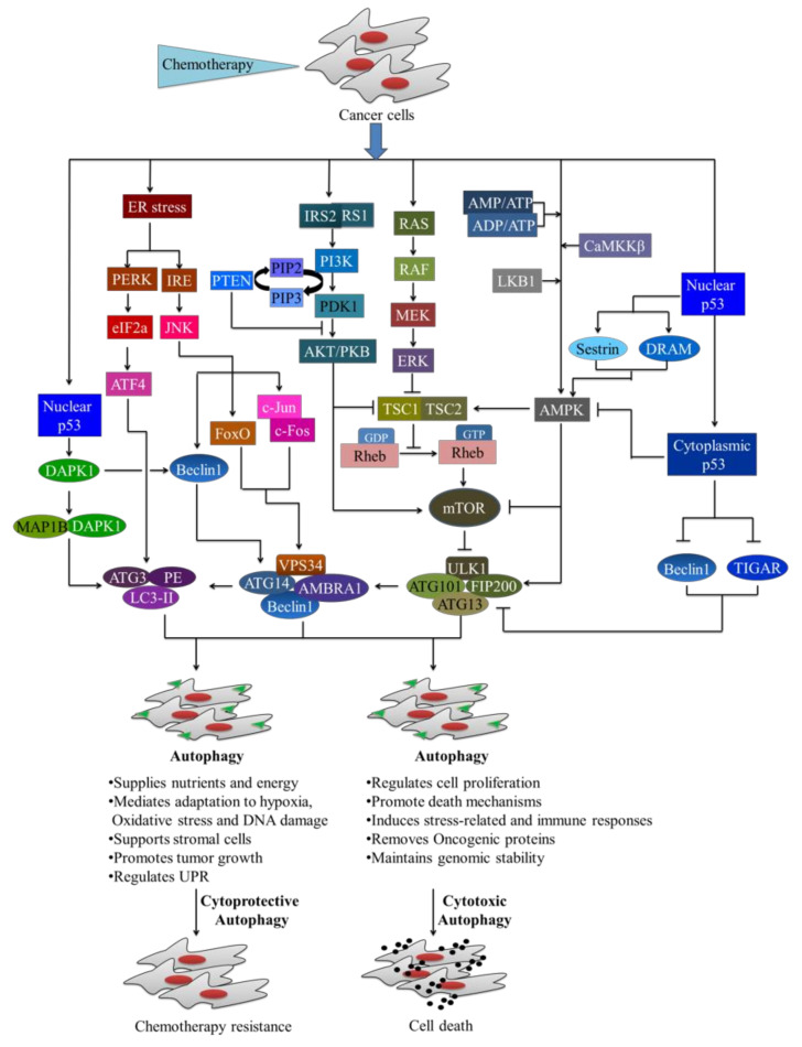
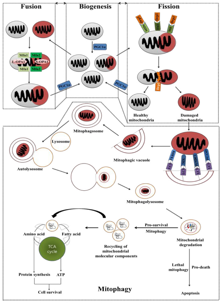
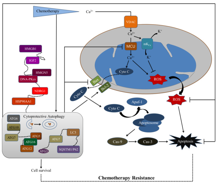
Cancer chemotherapy resistance is one of the most critical obstacles in cancer therapy. One of the well-known mechanisms of chemotherapy resistance is the change in the mitochondrial death pathways which occur when cells are under stressful situations, such as chemotherapy. Mitophagy, or mitochondrial selective autophagy, is critical for cell quality control because it can efficiently break down, remove, and recycle defective or damaged mitochondria. As cancer cells use mitophagy to rapidly sweep away damaged mitochondria in order to mediate their own drug resistance, it influences the efficacy of tumor chemotherapy as well as the degree of drug resistance. Yet despite the importance of mitochondria and mitophagy in chemotherapy resistance, little is known about the precise mechanisms involved. As a consequence, identifying potential therapeutic targets by analyzing the signal pathways that govern mitophagy has become a vital research goal. In this paper, we review recent advances in mitochondrial research, mitophagy control mechanisms, and their implications for our understanding of chemotherapy resistance.

Keywords: chemotherapy resistance; mitochondria; mitophagy.


Conflict of interest statement

The authors declare no conflict of interest.

Figures

Figure 1
Figure 1
Chemotherapy in development targeting mitochondrial components. The mitochondrial components are being targeted by chemotherapeutics that regulate metabolic activity, enabling for the selective killing of cancer cells for therapeutic benefit. To achieve therapeutic benefit, the calcium ion (Ca2+) channels or other ion complexes are predominantly targeted by chemotherapy, such as the voltage-dependent anion channel 1 (VDAC1), the mitochondrial calcium uniporter (MCU) complex, and the mitochondrial permeability transition pore complex (mPTPC), etc. Chemotherapy also targeted various mitochondrial enzymes or proteins, including mitochondrial ribosome and ribosomal proteins (MRP), DNA polymerase subunit gamma (PolG) and ATP synthase, etc. The mitochondrial deoxyribonucleic acid (mtDNA) and mitochondria-derived reactive oxygen species (mtROS) are considered to be potential mitochondrial components for chemotherapy. mPPT: mitochondrial permeability transition pore complex; ADP: adenosine diphosphate; ATP: adenosine triphosphate; mtDNA: mitochondrial deoxyribonucleic acid (DNA); mRNA: messenger ribonucleic acid; tRNA: transfer ribonucleic acid; VDAC1: voltage-dependent anion channel1; MCU: mitochondrial calcium uniporter; PolG: DNA polymerase gamma subunit.
Figure 2
Figure 2
Metabolic activity of normal and cancer cells. The cells use glucose as their primary metabolic substrate, which is broken down into pyruvate by glycolytic enzymes, which ultimately produces adenosine triphosphate (ATP) either by glycolysis or oxidative phosphorylation (OXPHOS). In normal cells, most pyruvate is transported into mitochondria for further oxidation to produce ATP. In cancer cells, most pyruvate is converted to lactate and transported out through the MCT transporter. GLUT: glucose transporter; MCT: monocarboxylate transporters.
Figure 3
Figure 3
Drugs targeting glycolysis and oxidative phosphorylation (OXPHOS) to sensitize chemotherapy resistance cancer cells. The cells use glucose as their primary metabolic substrate, which is broken down into pyruvate by glycolysis, which produces adenosine triphosphate (ATP), and nicotinamide adenine dinucleotide (NAD) plus hydrogen (H) (NADH) in the presence of various glycolysis enzymes, including hexokinase (HK), phosphofructokinase (PFK), and phosphoglycerate mutase (PGAM), etc. In normal cells, most pyruvate is transported into mitochondria for further oxidation, whereas in cancer cells most pyruvate is converted to lactate in the presence of lactate dehydrogenase (LDH). The tricarboxylic acid (TCA) cycle is fueled by acetyl-CoA, which produces NADH, flavin adenine dinucleotide (FADH2), ATP, and metabolic intermediates required for proliferation. The mitochondrial respiratory chain complex (RCC) or electron transport chains (ETC) are composed of complex I, complex II, complex III, complex IV and complex V (ATP synthase), that catalyzes the oxidation of reducing equivalents, primarily NADH and FADH2, in the IMM using the terminal electron acceptor oxygen (O2). The ETC receives NADH and FADH2 from the TCA cycle in the mitochondrial matrix, each of which provides a pair of electrons to the ETC via complexes I and II, respectively. The ETC complexes (complexes I–IV), the ATP synthase, the IMM, two diffusible electron carriers, and substrate transporters in IMM form the OXPHOS system. Ubiquinone (Q), which can be reduced to ubiquinol (QH2), is the product of electron transfer from complex I or II. Complex III oxidizes ubiquinol, allowing one electron to continue its journey via cytochrome c at a time. Cytochrome c transfers electrons to complex IV, where they are reduced to water by acting as a terminal electron acceptor. The reduction of O2 to H2O results in the pumping of protons (H+) to the IMS at complex IV. The re-entry of H+ into the matrix via complex V is related to the generation of ATP from ADP. OXPHOS: oxidative phosphorylation; GLUT1: glucose transporter 1; NADH: nicotinamide adenine dinucleotide (NAD) hydrogen; HK: hexokinase; PFK: phosphofructokinase; PGAM: phosphoglycerate mutase; LDH: lactate dehydrogenase; TCA: tricarboxylic acid; FADH2: adenine dinucleotide; RCC: respiratory chain complex; 2-DG: 2-deoxyglucose; 3-BP: 3-bromopyruvate; DHA: dehydroascorbic acid; MJ: methyl jasmonate; PKM: Pyruvate kinase M; PDH: pyruvate dehydrogenase; DCA: dichloroacetate; FCCP: carbonyl cyanide-p-trifluoromethoxyphenylhydrazone.
Figure 4
Figure 4
The process of autophagy. The process of autophagy is divided into three major stages: (i) initiation (formation of phagophore assembly site (PAS); (ii) nucleation, elongation and maturation (phagophore regulation and elongating into spherical autophagosomes); and (iii) fusion and degradation (autophagolysosome and autolysosome). mTOR: mammalian target of rapamycin; ULK1: unc-51-like kinase 1; ATGs: autophagy genes; FIP 200: focal adhesion kinase family interacting protein of 200 kDa; PI3K: phosphatidylinositol 3-kinase; PKB: phosphoinositide-dependent protein kinase B; VPS34: vacuolar protein sorting 34; AMBRA1: autophagy and beclin-1 regulator 1; LC3 I/II: light chain I and II; PE: phosphatidylethanolamine.
Figure 5
Figure 5
Possible pathways involved in autophagy-mediated cell survival or cell death. Chemotherapy can be activated cytotoxic or cytoprotective autophagy by a variety of signaling pathways; including the mTOR, phosphoinositide-dependent protein kinase B/AKT (PKB/AKT) pathway, AMP-activated protein kinase (AMPK), tumor suppressor p53 pathway, mitogen-activated protein kinase (MAPK) signaling, FOXO3A-PUMA signaling, and ER stress, etc. IRS1/2: insulin receptor substrate 1 and 2; PI3K; phosphatidylinositol 3-kinase; PDK1: phosphoinositide-dependent protein kinase 1; PTEN: phosphatase and tensin; PIP2: phosphatidylinositol-4,5-bisphosphate; PIP3: phosphatidylinositol-3,4,5-trisphosphate; PKB: phosphoinositide-dependent protein kinase B; RAS: rat sarcoma; RAF: rapidly accelerated fibrosarcoma; ERK: extracellular signal regulated kinases; MEK: mitogen-activated protein kinase/ERK kinase; TSC1/2: tuberous sclerosis complex 1 and 2; GDP: guanosine diphosphate; GT: guanosine triphosphate; mTOR: mammalian target of rapamycin; LC3-I/II: light chain 3 I and II; PERK: protein kinase R (PKR)-like endoplasmic reticulum kinase (PERK); eIF2: eukaryotic initiation factor 2; DAPK1: death-associated protein kinase 1; DRAM: damage regulated autophagy modulator; TIGAR: TP53-induced glycolysis and apoptosis regulator; LKB1: liver kinase B1; CaMKK: calcium/calmodulin-dependent protein kinase kinase; Rheb: ras homolog enriched in the brain; AMPK: AMP-activated protein kinase; ATG: autophagy gene.
Figure 6
Figure 6
The role of mitochondrial biogenesis and mitophagy in chemotherapy resistance. Mitochondrial biogenesis, fusion, fission, and mitophagy are cellular mechanisms that coordinately control mitochondrial content to maintain energy metabolism in response to cellular metabolic circumstances, stress, and other intracellular or external signals. The mitochondria increase their population by biogenesis process through utilizing fusion to fuse two neighboring mitochondria and fission to split two mitochondria from one, whereas the mitophagy (selective autophagic machinery) selectively removes damaged mitochondria to maintain cellular homeostasis. The pro-survival process of mitophagy utilized recycled mitochondrial components produced during the degradation process to use amino acids and fatty acids for protein synthesis and ATP generation, followed by cell survival (chemotherapy resistance). PGC1α: proliferator-activated receptor-gamma co-activator 1-alpha; OPA1: optic atrophy 1; Mnf1/2: mitofusin 1 and 2; Drp1: dynamin-related protein 1; FIS1: fission protein homolog 1; MFF: mitochondrial fission factor; MIDs: mitochondrial dynamics proteins; PINK1: PTEN-induced kinase 1; BNIP3: Bcl-2/adenovirus E1B 19 kDa interacting protein 3; FKBP8: FK506-binding protein 8; FUNDC1: FUN14 domain-containing protein 1; TCA: tricarboxylic acid.
Figure 7
Figure 7
Chemotherapy resistance, mitochondrial dysfunction and autophagic machinery. Mitochondria act as a powerful inducer of apoptosis via the intrinsic pathway, while mitochondrial calcium ion (Ca2+) regulates significant cell fate decisions like metabolism, growth, and death. The cytoprotective autophagy activated by chemotherapy through several autophagy regulating proteins that can effectively blocked Ca2+ channels on the outer mitochondrial membrane (OMM) and/or intramitochondrial membrane (IMM) to enter Ca2+. Inhibition of mitochondrial Ca2+ signaling can block the release of mitochondrial cytochrome c (Cyto C), followed by suppression of interaction between Cyto C and apoptosome, subsequently inhibit the apoptosis pathway. In addition, Ca2+ and Ca2+-activated potassium channel (mKca) has the ability to block the apoptosis pathway through the regulation of reactive oxygen species (ROS). Ca2+: calcium ion; HMGB1: high mobility group box 1; HMGN5: high-mobility group nucleosome-binding domain 5; HSP90AA1: heat shock protein 90AA1; IGF2: insulin growth factor 2; NDRG1: N-myc down-stream regulated gene 1; DNA-PKcs: DNA-dependent protein kinase catalytic subunit; mKca: Ca2+-activated potassium channel; Cyto C: cytochrome c; Bcl-2: B-cell lymphoma 2; Bax: BCL2-associated X; Apaf-1: apoptotic protease activating factor 1; Cas-9: caspase-9; Cas-3: caspase-3.

Similar articles

Cited by

References

    1. Yeldag G., Rice A., Del Rio Hernandez A. Chemoresistance and the Self-Maintaining Tumor Microenvironment. Cancers. 2018;10:471. doi: 10.3390/cancers10120471. - DOI - PMC - PubMed
    1. Assaraf Y.G., Brozovic A., Goncalves A.C., Jurkovicova D., Line A., Machuqueiro M., Saponara S., Sarmento-Ribeiro A.B., Xavier C.P.R., Vasconcelos M.H. The multi-factorial nature of clinical multidrug resistance in cancer. Drug Resist. Updates. 2019;46:100645. doi: 10.1016/j.drup.2019.100645. - DOI - PubMed
    1. Vaidya F.U., Sufiyan Chhipa A., Mishra V., Gupta V.K., Rawat S.G., Kumar A., Pathak C. Cancer Reports. Wiley Online Library; Hoboken, NJ, USA: 2020. Molecular and Cellular Paradigms of Multidrug Resistance in Cancer; p. e1291. - DOI - PMC - PubMed
    1. Bahar E., Kim J.Y., Yoon H. Chemotherapy Resistance Explained through Endoplasmic Reticulum Stress-Dependent Signaling. Cancers. 2019;11:338. doi: 10.3390/cancers11030338. - DOI - PMC - PubMed
    1. Bahar E., Kim J.Y., Kim H.S., Yoon H. Establishment of Acquired Cisplatin Resistance in Ovarian Cancer Cell Lines Characterized by Enriched Metastatic Properties with Increased Twist Expression. Int. J. Mol. Sci. 2020;21:7613. doi: 10.3390/ijms21207613. - DOI - PMC - PubMed