

Expression of PD-1, PD-L1 and PD-L2 in Lymphomas in Patients with Pre-Existing Rheumatic Diseases-A Possible Association with High Rheumatoid Arthritis Disease Activity
- PMID: 35326658
- PMCID: PMC8946311
- DOI: 10.3390/cancers14061509
Expression of PD-1, PD-L1 and PD-L2 in Lymphomas in Patients with Pre-Existing Rheumatic Diseases-A Possible Association with High Rheumatoid Arthritis Disease Activity
Abstract
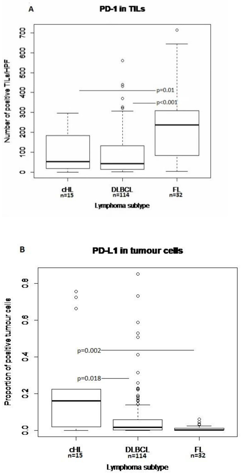
Current research seeks to identify subgroups of non-Hodgkin lymphoma (NHL) patients responsive to PD-1 blocking agents. Whether patients with pre-existing rheumatic diseases might constitute such a subgroup is unknown. We determined intratumoral expression of PD-1 and its ligands in lymphoma patients with pre-existing rheumatic diseases. We included 215 patients with rheumatoid arthritis (RA), systemic lupus erythematosus (SLE) or Sjögren's syndrome with subsequent lymphoma and 74 diffuse large B-cell lymphoma (DLBCL) controls without rheumatic disease. PD-1 and PD-ligand immunohistochemical markers were applied on tumor tissue microarrays. The number of PD-1+ tumor infiltrating leukocytes (TILs) and proportions of PD-L1+ and PD-L2+ tumor cells and TILs were calculated and correlated with clinical data. Expression of PD-L1 in tumor cells and TILs was highest in classical Hodgkin lymphoma and DLBCL. In DLBCLs, expression of PD-1 in TILs and PD-L1 in tumor cells was similar in RA, SLE and controls. In RA-DLBCL, high expression of PD-L1 in tumor cells was significantly more common in patients with the most severe RA disease and was associated with inferior overall survival in multivariable analysis.
Keywords: PD-1; PD-L1; immunohistochemistry; lymphoma; rheumatic disease.
Conflict of interest statement
The authors declare no competing financial interest. The funders had no role in the design of the study; in the collection, analyses, or interpretation of data, in the writing of the manuscript or in the decision to publish the results.
Figures


Similar articles
-
Expression of APRIL in diffuse large B cell lymphomas from patients with systemic lupus erythematosus and rheumatoid arthritis.J Rheumatol. 2011 Sep;38(9):1891-7. doi: 10.3899/jrheum.101190. Epub 2011 Jul 1. J Rheumatol. 2011. PMID: 21724695
-
Expression of programmed cell death 1/programmed cell death ligand 1 in the tumor microenvironments of primary gastrointestinal diffuse large B cell lymphomas.Pathol Res Pract. 2018 Apr;214(4):507-512. doi: 10.1016/j.prp.2018.03.001. Epub 2018 Mar 12. Pathol Res Pract. 2018. PMID: 29598887
-
PD-1/PD-L1 Pathway and Its Blockade in Patients with Classic Hodgkin Lymphoma and Non-Hodgkin Large-Cell Lymphomas.Curr Hematol Malig Rep. 2020 Aug;15(4):372-381. doi: 10.1007/s11899-020-00589-y. Curr Hematol Malig Rep. 2020. PMID: 32394185 Review.
-
Malignant lymphoma in patients with systemic rheumatic disease (rheumatoid arthritis, systemic lupus erythematosus, systemic sclerosis, and dermatomyositis): a clinicopathologic study of 24 Japanese cases.Int J Surg Pathol. 2006 Jan;14(1):43-8. doi: 10.1177/106689690601400108. Int J Surg Pathol. 2006. PMID: 16501834
-
Diagnostic utility of programmed cell death ligand 1 (clone SP142) immunohistochemistry for malignant lymphoma and lymphoproliferative disorders: A brief review.J Clin Exp Hematop. 2021 Dec 22;61(4):182-191. doi: 10.3960/jslrt.21003. Epub 2021 Sep 10. J Clin Exp Hematop. 2021. PMID: 34511582 Free PMC article. Review.
Cited by
-
The effect of combining PD-1 agonist and low-dose Interleukin-2 on treating systemic lupus erythematosus.Front Immunol. 2023 Mar 8;14:1111005. doi: 10.3389/fimmu.2023.1111005. eCollection 2023. Front Immunol. 2023. PMID: 36969198 Free PMC article. Review.
-
The susceptibility of single nucleotide polymorphisms located within co-stimulatory pathways to systemic lupus erythematosus.Front Immunol. 2024 Feb 1;14:1331796. doi: 10.3389/fimmu.2023.1331796. eCollection 2023. Front Immunol. 2024. PMID: 38361527 Free PMC article.
References
-
- Yamamoto R., Nishikori M., Kitawaki T., Sakai T., Hishizawa M., Tashima M., Kondo T., Ohmori K., Kurata M., Hayashi T., et al. PD-1-PD-1 ligand interaction contributes to immunosuppressive microenvironment of Hodgkin lymphoma. Blood. 2008;111:3220–3224. doi: 10.1182/blood-2007-05-085159. - DOI - PubMed
-
- Hollander P., Kamper P., Smedby K.E., Enblad G., Ludvigsen M., Mortensen J., Amini R.-M., Hamilton-Dutoit S., D’Amore F., Molin D., et al. High proportions of PD-1+ and PD-L1+ leukocytes in classical Hodgkin lymphoma microenvironment are associated with inferior outcome. Blood Adv. 2017;1:1427–1439. doi: 10.1182/bloodadvances.2017006346. - DOI - PMC - PubMed
-
- Roemer M.G., Advani R.H., Ligon A.H., Natkunam Y., Redd R.A., Homer H., Connelly C.F., Sun H.H., Daadi S.E., Freeman G.J., et al. PD-L1 and PD-L2 Genetic Alterations Define Classical Hodgkin Lymphoma and Predict Outcome. J. Clin. Oncol. 2016;34:2690–2697. doi: 10.1200/JCO.2016.66.4482. - DOI - PMC - PubMed
LinkOut - more resources
Full Text Sources
Research Materials
