. 2022 Mar 16;14(6):1517.
doi: 10.3390/cancers14061517.

Distinct Response of Circulating microRNAs to the Treatment of Pancreatic Cancer Xenografts with FGFR and ALK Kinase Inhibitors

Affiliations

Distinct Response of Circulating microRNAs to the Treatment of Pancreatic Cancer Xenografts with FGFR and ALK Kinase Inhibitors

Ivana Peran et al. Cancers (Basel). .

Abstract

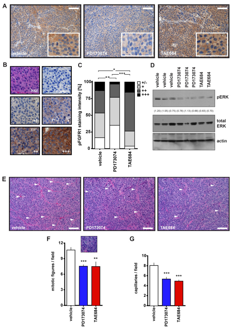
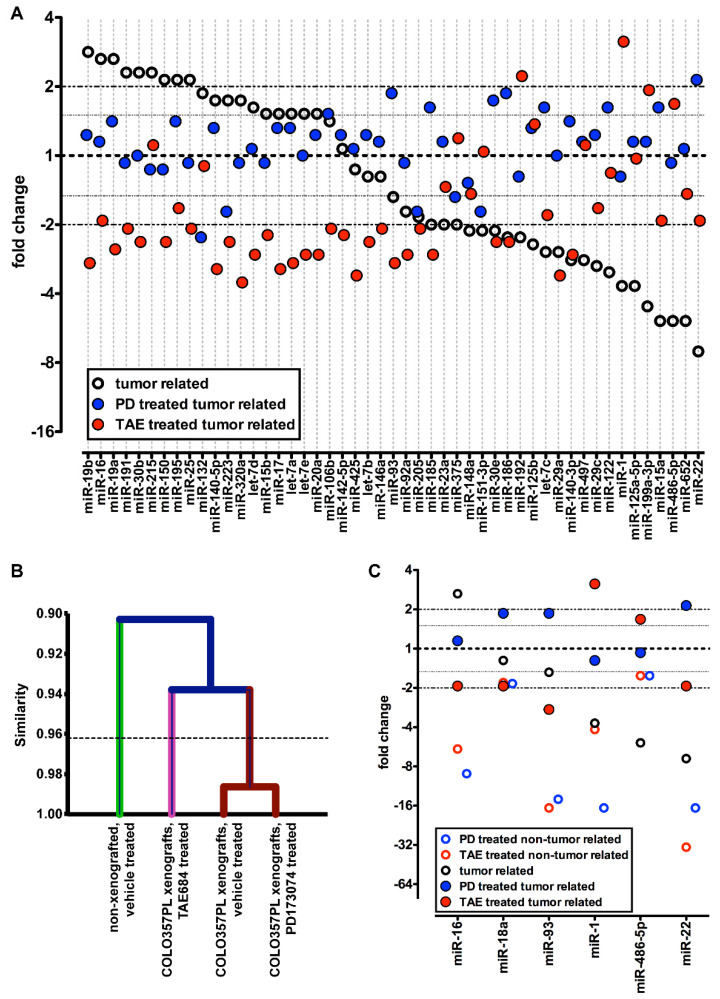
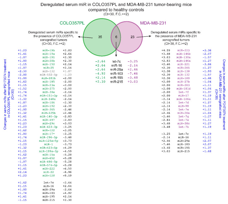
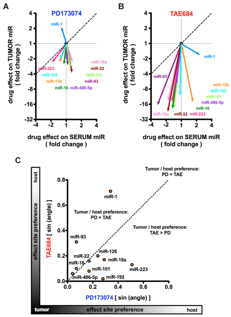
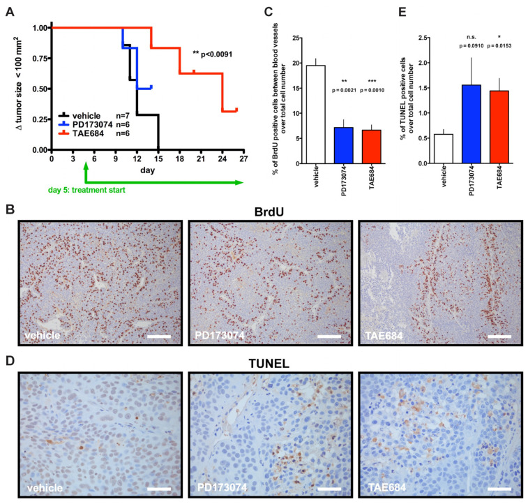
Pancreatic adenocarcinoma is typically detected at a late stage and thus shows only limited sensitivity to treatment, making it one of the deadliest malignancies. In this study, we evaluate changes in microRNA (miR) patterns in peripheral blood as a potential readout of treatment responses of pancreatic cancer to inhibitors that target tumor-stroma interactions. Mice with pancreatic cancer cell (COLO357PL) xenografts were treated with inhibitors of either fibroblast growth factor receptor kinase (FGFR; PD173074) or anaplastic lymphoma kinase receptor (ALK; TAE684). While both treatments inhibited tumor angiogenesis, signal transduction, and mitogenesis to a similar extent, they resulted in distinct changes in circulating miR signatures. Comparison of the miR pattern in the tumor versus that in circulation showed that the inhibitors can be distinguished by their differential impact on tumor-derived miRs as well as host-derived circulating miRs. Distinct signatures that include circulating miR-1 and miR-22 are associated with the efficacy of ALK and FGFR inhibition, respectively. We propose that monitoring changes in circulating miR profiles can provide an early signature of treatment response or resistance to pathway-targeted drugs, and thus provide a non-invasive measurement to rapidly assess the efficacy of candidate therapies.

Keywords: biomarker; circulating miR; miR; microRNA; pancreatic adenocarcinoma; pancreatic cancer; stroma; treatment response.


Conflict of interest statement

The authors declare no conflict of interest.

Figures

Figure 1
Figure 1
Response of COLO357PL cells to PD173074 or TAE684 in vitro. (A,B): Proliferation assays. COLO357PL cells were plated in the presence of drug in duplicates and measurements were made every 15 min. Cells were treated with PD173074 at 6 nM, 25 nM or 100 nM (A), or TAE684 at 2 nM, 8 nM or 32 nM (B). DMSO diluted in cell growth media served as control. Proliferation curves are shown as mean ± SEM. Two-way ANOVA was used as a statistical test and no significant p-values for treatment were observed. (C,D): Vindelov staining for cell cycle, and (E,F) Annexin V staining for apoptosis detection was analyzed by flow cytometry. Cells were treated for 48 h with PD173074 (100 nM) or TAE684 (32 nM). Vehicle treatment was DMSO diluted in cell growth media. Experiments were performed in duplicates. Mean ± SEM is shown. Unpaired two-tailed t-test was used and significant p-values for the comparison of kinase inhibitor treatment versus vehicle control are indicated. (G): Endothelial invasion assay. HUVECs form a stable monolayer prior to addition of drug +/− COLO357PL cells. The cell index was measured every 5 min for the first 6 h after addition of cancer cells. The experiment was performed in duplicates. Mean ± SEM is shown. Two-way ANOVA and p-values for treatment are indicated.
Figure 2
Figure 2
Effect of PD173074 and TAE684 7-day treatment on COLO357PL xenografts. (A): phospho-FGFR1 (pFGFR1) staining in representative tumor sections from different treatment groups (vehicle, PD173074 and TAE684), with zoomed inserts showing the localization of the staining. Scale bar, 0.1 mm. (B): Representative images of the pFGFR1 staining scale from negative to (+++) staining. (C): pFGFR1 staining intensity of the three treatment groups. * p < 0.05; ** p < 0.01; *** p < 0.0001; chi-square test. (D): Western blot for phospho- and total ERK of protein lysates from frozen tumor samples. (E): Representative H&E-stained tumor sections of the three treatment groups. Scale bar, 0.1 mm. (F,G): Ten pictures of different fields per tumor sample were analyzed for mitotic figures (F) and the number of capillaries (G). Mean ± SEM, ** p < 0.01; *** p ≤ 0.0002; unpaired two-tailed t-test. White arrowheads in panel (E) indicate capillaries. The insert in panel (F) shows an example of a mitotic figure. All the whole western blot figures can be found in the supplementary materials.
Figure 3
Figure 3
Serum miRs in nude mice with and without tumors and drug treatment. (A): Effect of tumor xenografts (black circles); fold changes of serum miRs in mice with COLO357PL tumors relative to controls without tumors. Effect of a 7-day drug treatment on serum miRs in COLO357PL tumor bearing mice (PD173074, blue symbols; TAE684, red symbols); fold changes in serum miRs after treatment relative to control treatment. qPCR was used to analyze 352 miRs, which were normalized for median Ct value. Fold changes are arranged by the expression of tumor-related miRs (black circles). Forty-nine serum miRs with Ct < 30 in every experimental group that were up- or downregulated ≥ two-fold in at least one of the comparisons are included. (B): Hierarchical clustering based on serum miR expression. The dotted line indicates p < 0.05. (C): Effect of PD173074 (blue circles) or TAE684 (red circles) treatment of mice without tumors. Six serum miRs were analyzed for expression in serum samples of nude mice without cancer that underwent short-term treatment. Data were normalized for U6 small nuclear RNA. Data from COLO357PL xenografted mice from panel (A) are included for comparison.
Figure 4
Figure 4
Venn diagram of serum miRs related to the presence of COLO357PL and MDA-MB-231 xenograft tumors, and serum miRs related to the short-term treatment with the FGFR inhibitor, PD173074. The expression levels of 352 serum miRs in COLO357PL (green) and 88 serum miRs in the MDA-MB-231 (magenta) xenograft mouse model were analyzed by qPCR and normalized for respective median Ct value. miRs with Ct < 30 that were changed by ≥2-fold are presented. Additionally, serum miRs changes after short-term treatment with PD173074 (blue) in both xenograft models are presented. Fold changes in serum miR expression after the PD173074 treatment are presented for every tumor-related miR with Ct < 30, as well as those that are ≥2-fold deregulated after PD173074 treatment.
Figure 5
Figure 5
Comparison of serum and tumor miR expression in COLO357PL xenograft mouse model. (A,B): PD173074 (A) or TAE684 (B) treatment-induced changes of miRs in serum and tumor samples relative to vehicle treatment. The vectors indicate the fold change of expression in the serum (x-axis) and tumor (y-axis) samples harvested at the same time. qPCR for miRs in serum samples were run in duplicates on previously pooled serum samples of mice that belonged to the same treatment group (n = 6 and 7 serum samples per treatment group); tumor samples were analyzed separately for miR expression (n = 7 and 8 tumors per treatment group). The dotted line indicates identical changes in tumor and serum. (C): Tumor or host (serum) preferential effect site for PD173074 versus TAE684 treatment. The sine of the direction of each miR change vector in panels (A,B) are plotted. The dotted line indicates identical preference of TAE and PD for the respective miR.
Figure 6
Figure 6
Effect of PD173074 and TAE684 treatment on COLO357PL tumor xenografts. (A): Kaplan–Meier plot of tumor growth >100 mm2 during the three-week treatment period. TAE684 vs. control; p-value by Gehan–Breslow–Wilcoxon test is indicated. PD173074 p > 0.05 vs. control. (B,C): BrdU staining for proliferating cells. Representative images of immunostaining are shown. Scale bar, 0.2 mm (B). Proliferating cells > 3 cell layers distant from blood vessels were quantified (C). A minimum of 22 images per group (total of 71) were analyzed. (D,E): TUNEL staining for apoptotic cells. Representative pictures are shown. Scale bar, 0.05 mm (D). Panels (BE): Vehicle treated mice, n = 4; drug treatment groups, n = 3 each; mean ± SEM; p-values from unpaired two-tailed t-test.

Similar articles

Cited by

References

    1. American Cancer Society . Cancer Facts & Figures 2020. American Cancer Society; Atlanta, GA, USA: 2020. pp. 1–76.
    1. Guturu P., Shah V., Urrutia R. Interplay of tumor microenvironment cell types with parenchymal cells in pancreatic cancer development and therapeutic implications. J. Gastrointest. Cancer. 2009;40:1–9. doi: 10.1007/s12029-009-9071-1. - DOI - PubMed
    1. Erkan M., Hausmann S., Michalski C.W., Fingerle A.A., Dobritz M., Kleeff J., Friess H. The role of stroma in pancreatic cancer: Diagnostic and therapeutic implications. Nat. Rev. Gastroenterol. Hepatol. 2012;9:454–467. doi: 10.1038/nrgastro.2012.115. - DOI - PubMed
    1. Rhim A.D., Oberstein P.E., Thomas D.H., Mirek E.T., Palermo C.F., Sastra S.A., Dekleva E.N., Saunders T., Becerra C.P., Tattersall I.W., et al. Stromal elements act to restrain, rather than support, pancreatic ductal adenocarcinoma. Cancer Cell. 2014;25:735–747. doi: 10.1016/j.ccr.2014.04.021. - DOI - PMC - PubMed
    1. Özdemir B.C., Pentcheva-Hoang T., Carstens J.L., Zheng X., Wu C.-C., Simpson T.R., Laklai H., Sugimoto H., Kahlert C., Novitskiy S.V., et al. Depletion of carcinoma-associated fibroblasts and fibrosis induces immunosuppression and accelerates pancreas cancer with reduced survival. Cancer Cell. 2014;25:719–734. doi: 10.1016/j.ccr.2014.04.005. - DOI - PMC - PubMed

LinkOut - more resources