

High-Mannose N-Glycans as Malignant Progression Markers in Early-Stage Colorectal Cancer
- PMID: 35326703
- PMCID: PMC8945895
- DOI: 10.3390/cancers14061552
High-Mannose N-Glycans as Malignant Progression Markers in Early-Stage Colorectal Cancer
Abstract
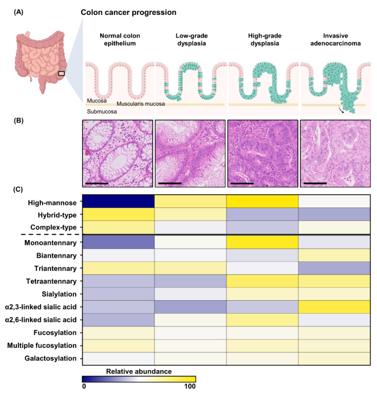
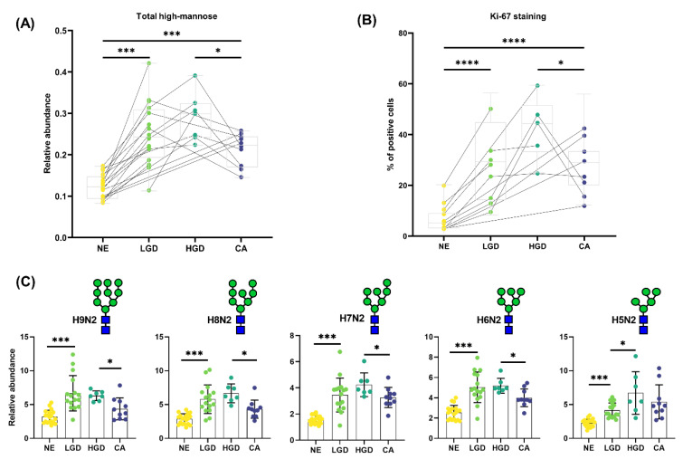
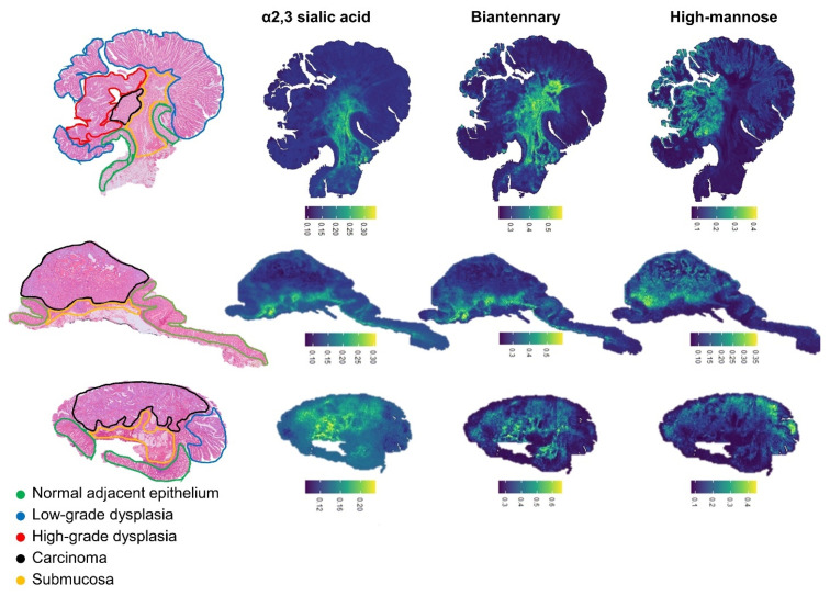
The increase incidence of early colorectal cancer (T1 CRC) last years is mainly due to the introduction of population-based screening for CRC. T1 CRC staging based on histological criteria remains challenging and there is high variability among pathologists in the scoring of these criteria. It is crucial to unravel the biology behind the progression of adenoma into T1 CRC. Glycomic studies have reported extensively on alterations of the N-glycomic pattern in CRC; therefore, investigating these alterations may reveal new insights into the development of T1 CRC. We used matrix-assisted laser desorption ionization (MALDI) mass spectrometry imaging (MSI) to spatially profile the N-glycan species in a cohort of pT1 CRC using archival formalin-fixed and paraffin-embedded (FFPE) material. To generate structural information on the observed N-glycans, CE-ESI-MS/MS was used in conjunction with MALDI-MSI. Relative intensities and glycosylation traits were calculated based on a panel of 58 N-glycans. Our analysis showed pronounced differences between normal epithelium, dysplastic, and carcinoma regions. High-mannose-type N-glycans were higher in the dysplastic region than in carcinoma, which correlates to increased proliferation of the cells. We observed changes in the cancer invasive front, including higher expression of α2,3-linked sialic acids which followed the glycosylation pattern of the carcinoma region.
Keywords: MALDI-MSI; N-glycosylation; colorectal cancer; early cancer progression; mass spectrometry imaging; molecular histology.
Conflict of interest statement
The authors declare no conflict of interest.
Figures
References
-
- Hari D.M., Leung A.M., Lee J.-H., Sim M.-S., Vuong B., Chiu C.G., Bilchik A.J. AJCC Cancer Staging Manual 7th edition criteria for colon cancer: Do the complex modifications improve prognostic assessment? J. Am. Coll. Surg. 2013;217:181–190. doi: 10.1016/j.jamcollsurg.2013.04.018. - DOI - PMC - PubMed
Grants and funding
LinkOut - more resources
Full Text Sources
