

Subsequent Ultrasonographic Non-Visualization of the Ovaries Is Hastened in Women with Only One Ovary Visualized Initially
- PMID: 35326911
- PMCID: PMC8955926
- DOI: 10.3390/healthcare10030433
Subsequent Ultrasonographic Non-Visualization of the Ovaries Is Hastened in Women with Only One Ovary Visualized Initially
Abstract
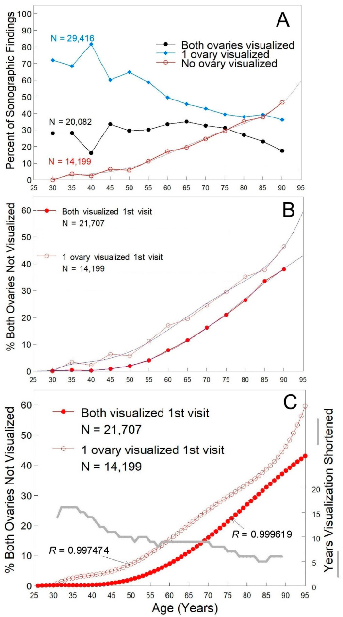
Because the effects of age, menopausal status, weight and body mass index (BMI) on ovarian detectability by transvaginal ultrasound (TVS) have not been established, we determined their contributions to TVS visualization of the ovaries when one or both ovaries are visualized on the first ultrasound exam. A total of 29,877 women that had both ovaries visualized on their first exam were followed over 202,639 prospective TVS exams and 9703 women that had only one ovary visualized on their first exam were followed over 63,702 ultrasonography exams. All images were reviewed by a physician. While non-visualization of both ovaries increased with age in women selected on the basis of the visualization of only one ovary on their first ultrasound exam, one or both ovaries could be visualized in two out of every three women at 80 years of age and more than 50% of women over 80 years of age. At each age, more non-visualizations were associated with women that had only one ovary visualized on their first visit. Having only one ovary visualized on the first exam advanced non-visualizations by an average of ~10 years across all ages and by >20 years in women under 40 years of age. Conclusions: Having only one ovary visualized on an initial ultrasound exam considerably hastens complete non-visualization for this population; however, in these women, ovaries can still be visualized well past menopause, and body habitus is not limiting to TVS ovarian imaging, thus TVS should be considered capable of capturing an ovarian image in two out of every three women at 80 years of age.
Keywords: BMI; age; body type; detection; menopausal status; ovary; transvaginal ultrasound; visualization; weight.
Conflict of interest statement
The authors declare no conflict of interest. The funders had no role in the design of the study; in the collection, analyses or interpretation of data; in the writing of the manuscript, or in the decision to publish the results.
Figures

Similar articles
-
Ultrasonographic Visualization of the Ovaries to Detect Ovarian Cancer According to Age, Menopausal Status and Body Type.Diagnostics (Basel). 2022 Jan 6;12(1):128. doi: 10.3390/diagnostics12010128. Diagnostics (Basel). 2022. PMID: 35054294 Free PMC article.
-
Factors affecting visualization of postmenopausal ovaries: descriptive study from the multicenter United Kingdom Collaborative Trial of Ovarian Cancer Screening (UKCTOCS).Ultrasound Obstet Gynecol. 2013 Oct;42(4):472-7. doi: 10.1002/uog.12447. Ultrasound Obstet Gynecol. 2013. PMID: 23456790 Clinical Trial.
-
Non-visualization of the ovary on CT or ultrasound in the ED setting: utility of immediate follow-up imaging.Abdom Radiol (NY). 2018 Sep;43(9):2467-2473. doi: 10.1007/s00261-017-1438-3. Abdom Radiol (NY). 2018. PMID: 29230555
-
Ovarian cancer screening.Cancer. 1995 Nov 15;76(10 Suppl):2086-91. doi: 10.1002/1097-0142(19951115)76:10+<2086::aid-cncr2820761330>3.0.co;2-l. Cancer. 1995. PMID: 8635005 Review.
-
Androgens in Menopausal Women: Not Only Polycystic Ovary Syndrome.Front Horm Res. 2019;53:135-161. doi: 10.1159/000494909. Epub 2019 Sep 9. Front Horm Res. 2019. PMID: 31499509 Review.
References
-
- Ovarian Cancer Including Fallopian Tube Cancer and Primary Peritoneal Cancer. [(accessed on 22 February 2022)]. Available online: https://www.nccn.org/professionals/physician_gls/pdf/ovarian.pdf.
-
- Jacobs I., Gentry-Maharaj A., Burnell M., Manchanda R., Singh N., Sharma A., Ryan A., Seif M.W., Amso N.N., Turner G., et al. Sensitivity of transvaginal ultrasound screening for endometrial cancer in postmenopausal women: A case-control study within the UKCTOCS cohort. Lancet Oncol. 2011;12:38–48. doi: 10.1016/S1470-2045(10)70268-0. - DOI - PubMed
-
- van Nagell J., Burgess B., Miller R., Baldwin L., DeSimone C., Ueland F., Huang B., Chen Q., Kryscio R., Pavlik E. Survival of women with type I and II epithelial ovarian cancer detected by ultrasound screening. Obstet. Gynecol. 2018;132:1091–1100. doi: 10.1097/AOG.0000000000002921. - DOI - PMC - PubMed
-
- Carter J., Fowler J., Carson L., Carlson J., Twiggs L.B. How accurate is the pelvic examination as compared to transvaginal sonography? A prospective, comparative study. J. Reprod. Med. 1994;39:32–34. - PubMed
Grants and funding
LinkOut - more resources
Full Text Sources
Miscellaneous
