. 2022 Mar 10;12(6):696.
doi: 10.3390/ani12060696.

A New Disease Caused by an Unidentified Etiological Agent Affects European Salamanders

Affiliations

A New Disease Caused by an Unidentified Etiological Agent Affects European Salamanders

Raoul Manenti et al. Animals (Basel). .

Abstract

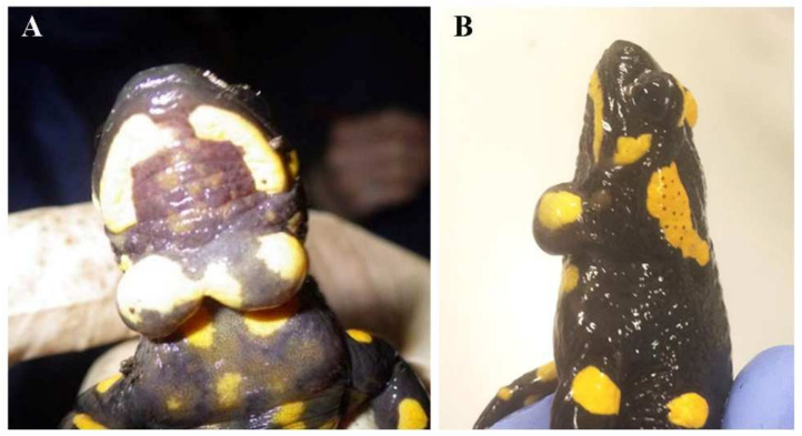
New pathologies are causing dramatic declines and extinctions of multiple amphibian species. In 2013, in one fire salamander population of Northern Italy, we found individuals with undescribed cysts at the throat level, a malady whose existence has not previously been reported in amphibians. With the aim of describing this novel disease, we performed repeated field surveys to assess the frequency of affected salamanders from 2014 to 2020, and integrated morphological, histological, and molecular analyses to identify the pathogen. The novel disease affected up to 22% of salamanders of the study population and started spreading to nearby populations. Cysts are formed by mucus surrounding protist-like cells about 30 µm long, characterized by numerous cilia/undulipodia. Morphological and genetic analyses did not yield a clear match with described organisms. The existence of this pathogen calls for the implementation of biosecurity protocols and more studies on the dynamics of transmission and the impact on wild populations.

Keywords: Mesomycetozoea; Salamandra salamandra; amphibians; fire salamander; pathogenic; protist.


Conflict of interest statement

The authors declare no conflict of interest.

Figures

Figure 1
Figure 1
Salamander cysts. Ventral (A) and lateral (B) view of a male Salamandra salamandra with two turgid cysts occurring in a median position at the throat level.
Figure 2
Figure 2
Location of the study area. (A) location and area of the transects monitored, identified by the letter T; “Guercio” identifies the main locality on the regional protected area named “Riserva del Guercio” in which the pathogen was first detected. “Olgelasca” identifies the adjacent locality in which the pathogen appeared in 2016. (B) picture of the protected area between transect 1 and transect 2.
Figure 3
Figure 3
Morphological analyses. (AC) Cyst cell culture in which peculiar ciliated cells are observable (arrow). These protist-like cells displayed heterogeneous cytoplast and their cell membrane was covered by numerous motile cilium-like structures (B,C). (DF) Histological analysis revealed that the cyst masses consisted of a thin capsule of connective tissue encircling a central area of mucus material in which numerous granulocytes, plasma cells, and lymphocytes were detected (D,E). Among these, ciliated protist-like cells were present trapped in the mucus (F); asterisks identify trapped cells. Scale bars: (A) 20 µm; (B,C) 10 µm; (D) 250 µm; (E,F) 50 µm.
Figure 4
Figure 4
Electron microscopy analyses. (AE) Transmission electron microscopy of the pathogen. In cyst mucus, numerous protist-like cells (A) with long undulipodia were found. At higher magnification (B), electron dense and transparent vacuoles were observed as well as undulipodium rootlets regularly distributed along cytoplasm periphery (C,D). Mitochondria with peculiar cristae were detected (E). (FI) Scanning electron microscopy. Trapped inside mucus niches, protist-like cells were observed (F,G). Their main feature was the numerous and long cilia/undulipodia which covered the cell membrane (H,I). Scale bars: (A) 2 µm; (BD) 1 µm; (E) 500 nm; (F) 10 µm; (G) 5 µm; (H) 2 µm; (I) 1 µm.

Similar articles

Cited by

References

    1. Cleaveland S., Haydon D.T., Taylor L. Overviews of pathogen emergence: Which pathogens emerge, when and why? Curr. Top. Microbiol. Immunol. 2007;315:85–111. doi: 10.1007/978-3-540-70962-6_5. - DOI - PMC - PubMed
    1. Lu M.M., Wang X.D., Ye H., Wang H.M., Qiu S., Zhang H.M., Liu Y., Luo J.H., Feng J. Does public fear that bats spread COVID-19 jeopardize bat conservation? Biol. Conserv. 2021;254:9. doi: 10.1016/j.biocon.2021.108952. - DOI - PMC - PubMed
    1. Maurin M., Fenollar F., Mediannikov O., Davoust B., Devaux C., Raoult D. Current Status of Putative Animal Sources of SARS-CoV-2 Infection in Humans: Wildlife, Domestic Animals and Pets. Microorganisms. 2021;9:868. doi: 10.3390/microorganisms9040868. - DOI - PMC - PubMed
    1. Schrag S.J., Wiener P. Emerging infectious disease: What are the relative roles of ecology and evolution? Trends Ecol. Evol. 1995;10:319–324. doi: 10.1016/S0169-5347(00)89118-1. - DOI - PubMed
    1. Murray A.G., Smith R.J., Stagg R.M. Shipping and the spread of infectious salmon anemia in Scottish aquaculture. Emerg. Infect. Dis. 2002;8:1–5. doi: 10.3201/eid0801.010144. - DOI - PMC - PubMed