

Influence of Obesity on Histological Tissue Structure of the Cardiovascular System in Horses
- PMID: 35327129
- PMCID: PMC8944453
- DOI: 10.3390/ani12060732
Influence of Obesity on Histological Tissue Structure of the Cardiovascular System in Horses
Abstract
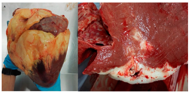
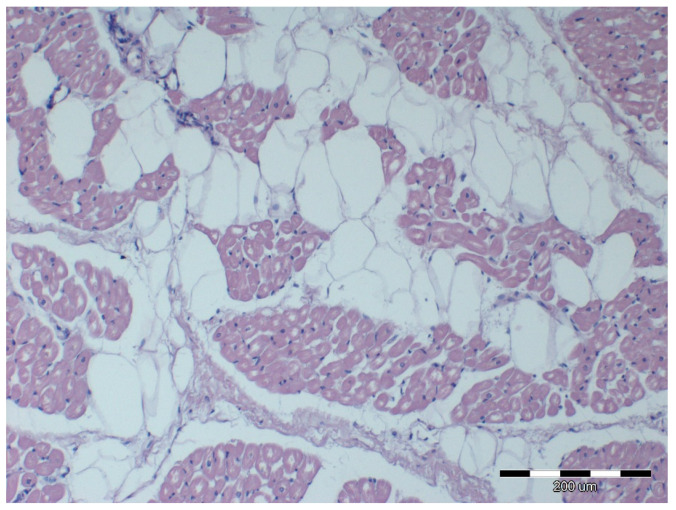
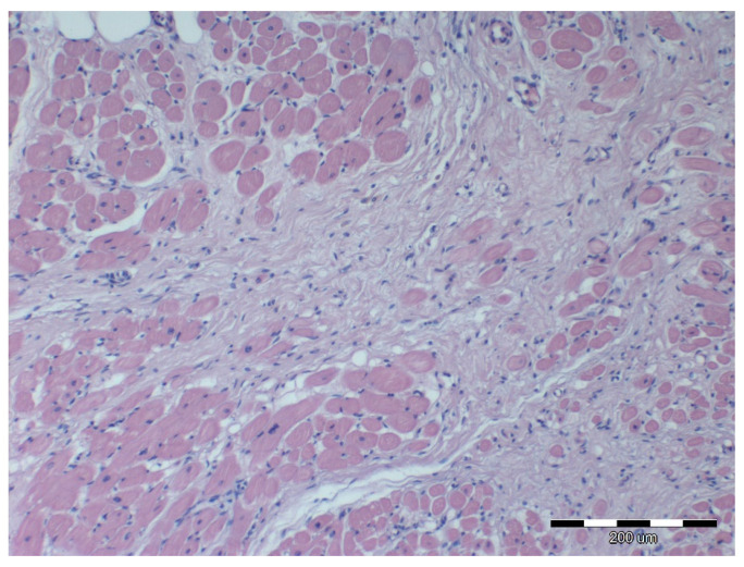
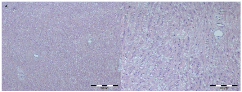
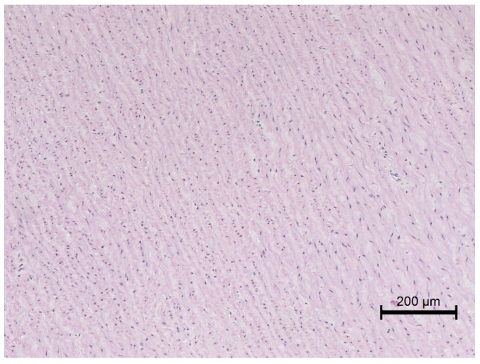
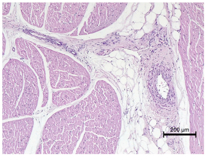
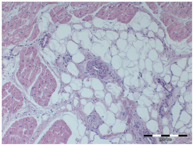
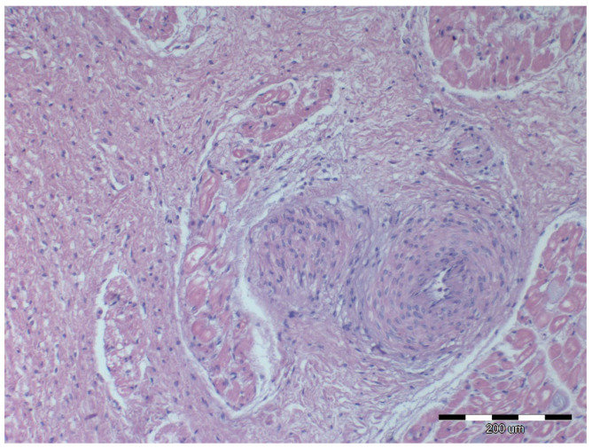
It has been well established that obesity in horses can have a negative impact on their health, including endocrine disturbances. In humans, it is well known that obesity leads to structural and functional changes of the cardiovascular system. The aim of the study was to assess the impact of obesity on the histological structure of the myocardial tissue, as well as great and peripheral arteries in horses. The heart, arteries (aorta, pulmonary artery and palmar arteries) specimens from 7 horses with normal BCS (4-5/9) and 12 extremely obese (BCS 9/9) draft slaughter horses were obtained for histopathological evaluation. The heart tissue and great arteries showed more intense disturbances in the architecture and vacuolization in the aorta in obese horses as compared to the healthy group. The intima in the pulmonary artery, coronary arteries and palmar arteries was thicker in the obese, rather than healthy horses. The palmar arteries in obese horses had a larger lumen diameter and the lumen-to-total diameter ratio as compared to the control group. The presented study showed a significant effect of obesity on the heart as well as the central and peripheral vessels in horses. This forms the basis for a deeper reflection on the impact of obesity on the equine body.
Keywords: equine; heart; leptin; overweight; vessels.
Conflict of interest statement
The authors declare that the research was conducted in the absence of any commercial or financial relationships that could be construed as a potential conflict of interest.
Figures

Similar articles
-
[The status of large vessels in overweight and obesity according to ultrasound findings].Ter Arkh. 2016;88(4):29-34. doi: 10.17116/terarkh201688429-34. Ter Arkh. 2016. PMID: 27070160 Russian.
-
Body condition score, morphometric measurements and estimation of body weight in mature Icelandic horses in Denmark.Acta Vet Scand. 2016 Oct 20;58(Suppl 1):59. doi: 10.1186/s13028-016-0240-5. Acta Vet Scand. 2016. PMID: 27766968 Free PMC article.
-
Tall fescue ergot alkaloids are vasoactive in equine vasculature.J Anim Sci. 2017 Nov;95(11):5151-5160. doi: 10.2527/jas2017.1852. J Anim Sci. 2017. PMID: 29293720 Free PMC article.
-
Assessment of peripheral blood neutrophil respiratory burst, phagocytosis and apoptosis in obese non-insulin dysregulated horses.Res Vet Sci. 2020 Oct;132:127-132. doi: 10.1016/j.rvsc.2020.06.013. Epub 2020 Jun 12. Res Vet Sci. 2020. PMID: 32563928
-
The equine metabolic syndrome peripheral Cushing's syndrome.Vet Clin North Am Equine Pract. 2002 Aug;18(2):271-93. doi: 10.1016/s0749-0739(02)00006-8. Vet Clin North Am Equine Pract. 2002. PMID: 15635908 Review.
Cited by
-
Impacts of Adiposity on Exercise Performance in Horses.Animals (Basel). 2023 Feb 14;13(4):666. doi: 10.3390/ani13040666. Animals (Basel). 2023. PMID: 36830453 Free PMC article. Review.
-
Obesity during Pregnancy in the Horse: Effect on Term Placental Structure and Gene Expression, as Well as Colostrum and Milk Fatty Acid Concentration.Vet Sci. 2023 Dec 4;10(12):691. doi: 10.3390/vetsci10120691. Vet Sci. 2023. PMID: 38133242 Free PMC article.
-
Confidence does not mediate a relationship between owner experience and likelihood of using weight management approaches for native ponies.PLoS One. 2023 Oct 12;18(10):e0292886. doi: 10.1371/journal.pone.0292886. eCollection 2023. PLoS One. 2023. PMID: 37824555 Free PMC article.
-
Effect of Exercise Conditioning on Countering the Effects of Obesity and Insulin Resistance in Horses-A Review.Animals (Basel). 2024 Feb 26;14(5):727. doi: 10.3390/ani14050727. Animals (Basel). 2024. PMID: 38473112 Free PMC article. Review.
References
LinkOut - more resources
Full Text Sources
