. 2022 Mar 18;11(6):863.
doi: 10.3390/foods11060863.

Research on Rapid Detection Technology for β2-Agonists: Multi-Residue Fluorescence Immunochromatography Based on Dimeric Artificial Antigen

Affiliations

Research on Rapid Detection Technology for β2-Agonists: Multi-Residue Fluorescence Immunochromatography Based on Dimeric Artificial Antigen

Miaomiao Liu et al. Foods. .

Abstract

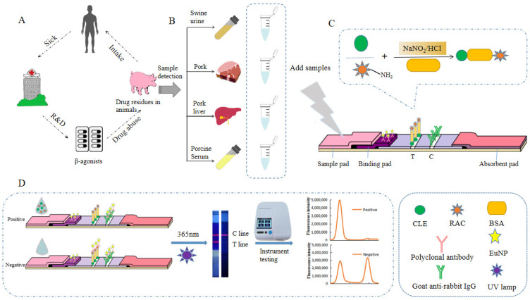
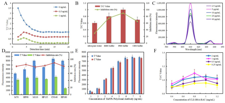
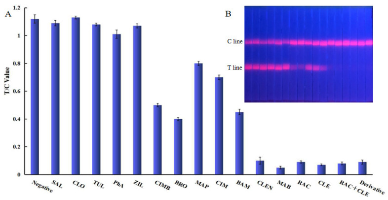
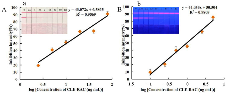
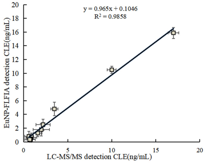
To detect two types of β2-agonist residues at the same time, we coupled two haptens of clenbuterol (CLE) and ractopamine (RAC) to the same carrier protein through diazotization to prepare dimeric artificial antigen, and a fluorescent lateral flow immunoassay method based on europium nanoparticles (EuNP-FLFIA) was established by combining polyclonal antibodies with europium nanoparticles to form probes. Under optimized conditions, the EuNP-FLFIA could simultaneously detect eight aniline-type and one phenol-type β2-agonists, and the limits of detection (LOD) were 0.11−0.19 ng/mL and 0.12 ng/mL, respectively. The recovery rate of this method was 84.00−114.00%. This method was verified by liquid chromatography−tandem mass spectrometry (LC-MS/MS), and the test results were consistent (R2 > 0.98). Therefore, the method established in this study could be used as a high-throughput screening for the efficient and sensitive detection of β2-agonists in food.

Keywords: dimeric artificial antigen; europium nanoparticles; fluorescent lateral flow immunoassay; multi-residue analysis; β2-agonists.


Conflict of interest statement

The authors declare no conflict of interest.

Figures

Figure 1
Figure 1
The EuNP-FLFIA detection principle. (A) The effects of β2-agonist abuse on the human body through the food chain. (B) Detection of actual samples. (C) Synthesis of dimeric artificial antigen. (D) The testing process and test results of the EuNP-FLFIA reader test results.
Figure 2
Figure 2
Optimization of the EuNP-FLFIA assay. (A) The relationship between detection time and the T/C value. The sample concentration was 0, 0.5, and 1 ng/mL, respectively. (B) Effect of buffers on the T/C value. Buffer types: ultra-pure water, BBS buffer, PBS buffer, and CBS buffer. (C) Effect of different EuNP polyclonal antibody probe concentrations on the fluorescence intensity of the mixed solutions. (D) The fluorescence intensity values and inhibition intensity are shown by using different nitrocellulose membranes. Nitrocellulose membrane types: M70, HF90, M110, HF135, CN140, and HF180. (E) Effect of EuNP polyclonal antibody probe concentrations on the fluorescence intensity. (F) Influence of various CLE-BSA-RAC concentrations and EuNP polyclonal antibody probe concentrations on the T/C value.
Figure 3
Figure 3
Specific analysis of EuNP-FLFIA. (A) Specific evaluation results (from left to right are negative: SAL, CLO, TUL, PhA, ZIL, CIMB, BRO, MAP, CIM, BAM, CLEN, MAB, RAC, CLE, RAC+CLE, and Derivative). (B) T/C value data analysis showing the extent of the cross-reactivity of CLE-RAC with other β2-agonists.
Figure 4
Figure 4
Detection in EuNP-FLFIA and CG-LFIA. (A). The standard curve for CLE+RAC (1:1) by CG-LFIA. a. Physical diagram of CG-LFIA for CLE-RAC sensitivity detection. (B). The standard curve for CLE+RAC (1:1) by EuNP-LFIA. b. Physical diagram of EuNP-FLFIA for CLE-RAC sensitivity detection.
Figure 5
Figure 5
EuNP-FLFIA and LC-MS/MS comparison test results.

Similar articles

Cited by

References

    1. Flavie O.B.E., Wang H.V. In vitro Beta-agonist drugs modulate the proliferation and differentiation of skeletal muscle cells. Biochem. Biophys. Rep. 2021;26:101019. doi: 10.1016/j.bbrep.2021.101019. - DOI - PMC - PubMed
    1. Mastrianni K.R., Metavarayuth K., Brewer W.E., Wang Q. Analysis of 10 β-agonists in pork meat using automated dispersive pipette extraction and LC-MS/MS. J. Chromatogr. B. 2018;1084:64–68. doi: 10.1016/j.jchromb.2018.03.026. - DOI - PubMed
    1. Kaufmann A., Widmer M., Maden K., Butcher P., Walker S. High resolution mass spectrometry-based detection and quantification of β-agonists at relevant trace levels in a variety of animal-based food matrices. Food Addit. Contam. Part A Chem. Anal. Control. Expo. Risk Assess. 2021;38:1350–1363. doi: 10.1080/19440049.2021.1922759. - DOI - PubMed
    1. Kuiper H.A., Noordam M.Y., van Dooren-Flipsen M.M.H., Schilt R., Roos A.H. Illegal use of beta-adrenergic agonists: European Community. J. Anim. Sci. 1998;76:195–207. doi: 10.2527/1998.761195x. - DOI - PubMed
    1. Mitchell G.A., Dunnavan G. Illegal use of beta-adrenergic agonists in the United States. J. Anim. Sci. 1998;76:208–211. doi: 10.2527/1998.761208x. - DOI - PubMed

LinkOut - more resources