. 2022 Feb 23;10(3):527.
doi: 10.3390/biomedicines10030527.

Fibrin, Bone Marrow Cells and Macrophages Interactively Modulate Cardiomyoblast Fate

Affiliations

Fibrin, Bone Marrow Cells and Macrophages Interactively Modulate Cardiomyoblast Fate

Inês Borrego et al. Biomedicines. .

Abstract

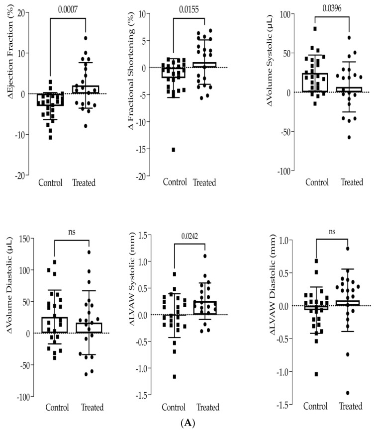
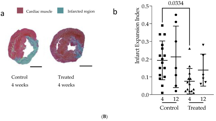
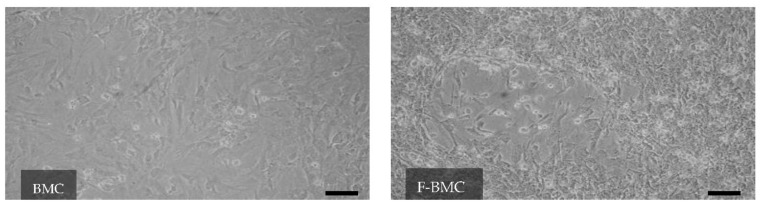
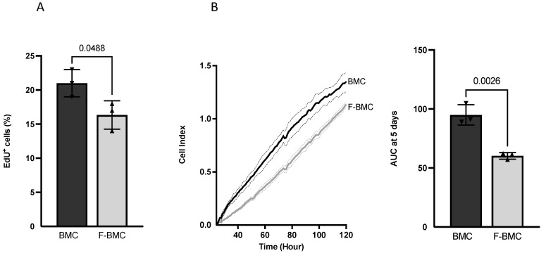
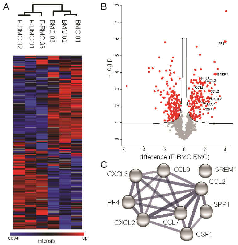
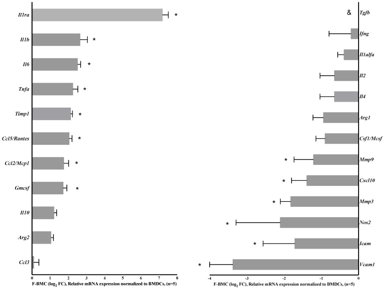
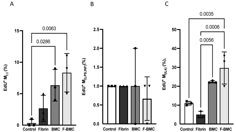
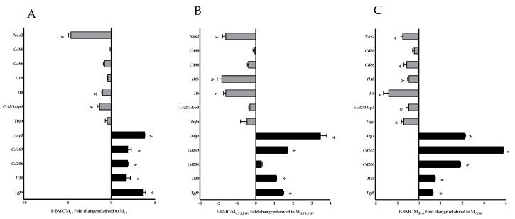
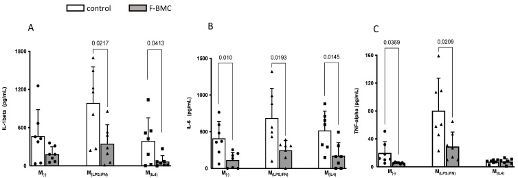
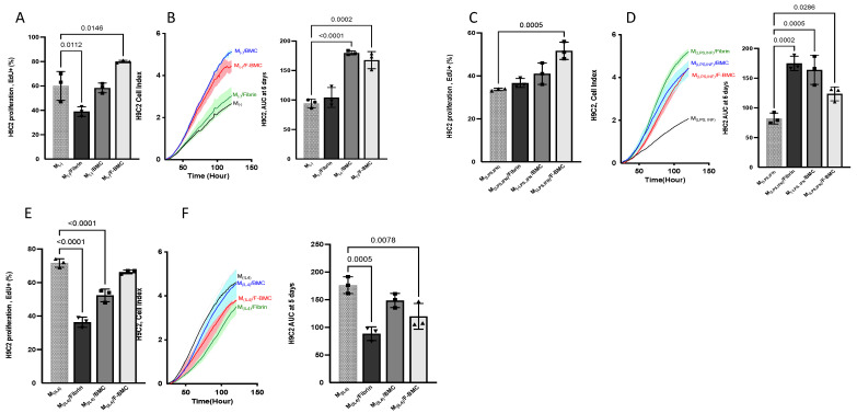
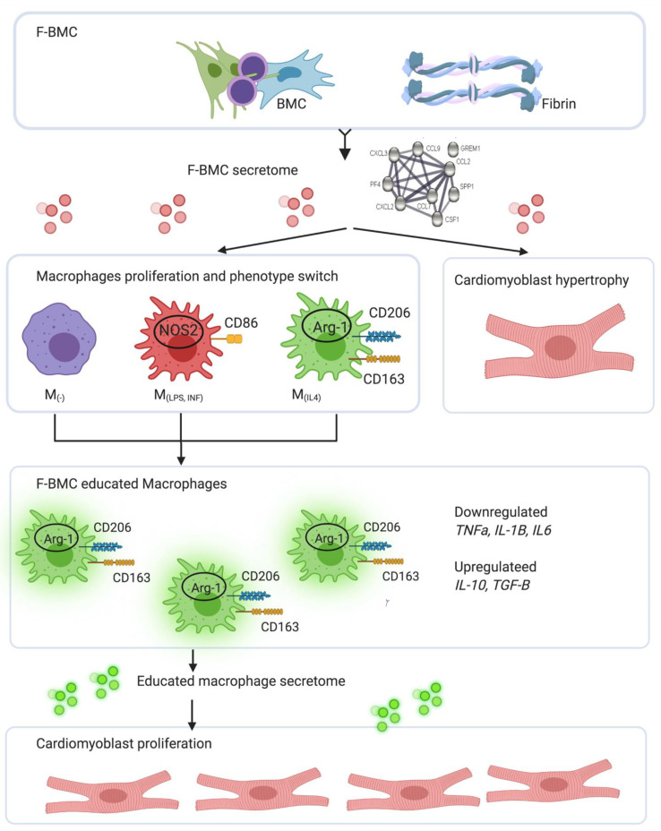
Interactions between macrophages, cardiac cells and the extracellular matrix are crucial for cardiac repair following myocardial infarction (MI). We hypothesized that cell-based treatments might modulate these interactions. After validating that bone marrow cells (BMC) associated with fibrin lowered the infarct extent and improved cardiac function, we interrogated the influence of fibrin, as a biologically active scaffold, on the secretome of BMC and the impact of their association on macrophage fate and cardiomyoblast proliferation. In vitro, BMC were primed with fibrin (F-BMC). RT-PCR and proteomic analyses showed that fibrin profoundly influenced the gene expression and the secretome of BMCs. Consequently, the secretome of F-BMC increased the spreading of cardiomyoblasts and showed an alleviated immunomodulatory capacity. Indeed, the proliferation of anti-inflammatory macrophages was augmented, and the phenotype of pro-inflammatory switched as shown by downregulated Nos2, Il6 and IL1b and upregulated Arg1, CD163, Tgfb and IL10. Interestingly, the secretome of F-BMC educated-macrophages stimulated the incorporation of EdU in cardiomyoblasts. In conclusion, our study provides evidence that BMC/fibrin-based treatment improved cardiac structure and function following MI. In vitro proofs-of-concept reveal that the F-BMC secretome increases cardiac cell size and promotes an anti-inflammatory response. Thenceforward, the F-BMC educated macrophages sequentially stimulated cardiac cell proliferation.

Keywords: cell communication; cell priming; fibrin; inflammation; macrophages; secretome.


Conflict of interest statement

The authors declare no conflict of interest.

Figures

Figure 1
Figure 1
(A) Fibrin and BMC treatment reduces systolic heart function loss. Cardiac function was assessed by high-resolution echocardiography. The changes (Δ) in ejection fraction, fractional shortening, left ventricle (LV) volumes and LV wall thickness (LVAW) were calculated as the difference between 4 weeks post- and pre-treatment. The control group is untreated (sham) infarcted animals (n = 23); the treated group is animals treated with epicardial fibrin and BMC (n = 20). (B) Fibrin and BMC treatment reduces fibrotic scar. (a) Representative Masson–Goldner trichrome stained heart cross-sections from treated and control (sham) animals, 4 weeks post-treatment. The fibrotic scar tissue is in blue; in red is the remote tissue. Scale bars indicate 3 mm. (b) Infarct Expansion Index was measured on histologic sections 4 weeks or 12 weeks post-treatment. A total of 5 to 6 cross-sections from systematic sampling of the whole heart were averaged for each animal. Each point represents one animal. The control group is infarcted animals that received sham treatment. The treated group is animals treated with epicardial fibrin and BMC. The values are shown as mean ± SD.
Figure 1
Figure 1
(A) Fibrin and BMC treatment reduces systolic heart function loss. Cardiac function was assessed by high-resolution echocardiography. The changes (Δ) in ejection fraction, fractional shortening, left ventricle (LV) volumes and LV wall thickness (LVAW) were calculated as the difference between 4 weeks post- and pre-treatment. The control group is untreated (sham) infarcted animals (n = 23); the treated group is animals treated with epicardial fibrin and BMC (n = 20). (B) Fibrin and BMC treatment reduces fibrotic scar. (a) Representative Masson–Goldner trichrome stained heart cross-sections from treated and control (sham) animals, 4 weeks post-treatment. The fibrotic scar tissue is in blue; in red is the remote tissue. Scale bars indicate 3 mm. (b) Infarct Expansion Index was measured on histologic sections 4 weeks or 12 weeks post-treatment. A total of 5 to 6 cross-sections from systematic sampling of the whole heart were averaged for each animal. Each point represents one animal. The control group is infarcted animals that received sham treatment. The treated group is animals treated with epicardial fibrin and BMC. The values are shown as mean ± SD.
Figure 2
Figure 2
Fibrin modulates the BMC morphology: Representative pictures of BMC cultured with fibrin (F-BMC) or without (BMC) showing a heterogeneous population of cells with different morphology and spreading. Scale bar = 100 μm.
Figure 3
Figure 3
Fibrin modulates BMC proliferation: BMC were cultured with (F-BMC) or without fibrin (BMC). Cell growth was assessed by (A) EdU incorporation for 48 h and by (B) RTCA: Cell index was measured over 120 h, plotted, and the AUC calculated. The values are shown as mean ± SD; n = 3 biologically independent samples of pools of the BMC of 3 rats i.
Figure 4
Figure 4
Expression proteomic analyses of BMCs cultured with and without a fibrin-based substrate. (A) Hierarchical clustering of protein abundances using log2 transformed and z-normalized LFQ intensities indicates global alterations of CM due to culture conditions. Grey squares indicate proteins not detected in respective samples. (B) Volcano plot analysis highlights significantly altered proteins due to culture condition in red (FDR < 0.05). The black line indicates S0 of 0.1. (C) Eight cytokines known to interact based on STRING DB are significantly downregulated in BMCs cultured with the fibrin-based substrate [29]. The thickness of edges indicates the confidence of data support.
Figure 5
Figure 5
Fibrin modulates BMC gene expression. Real-time PCR measurement of selected genes presented as fold change (FC). n = 5 biologically independent samples of cell pools from 3 rats. * p < 0.05, shows the statistical significance between differential gene expression of F-BMC related to BMC assessed by one-way ANOVA and Dunnett’s test.
Figure 6
Figure 6
F-BMC and BMC conditioned media modulate macrophage proliferation. The proliferation of the different macrophages phenotypes was assessed by EdU incorporation. (A) M(−), (B) M(LPS,IFN), and (C) M(IL-4) macrophages were cultured with unconditioned medium (Control) or conditioned media from with Fibrin, F-BMC and BMC. The values shown are mean ± SD. n = 3 biologically independent pools of macrophages from 3 animals, therefore, in total, 9 animals per group.
Figure 7
Figure 7
F-BMC conditioned medium modulates the macrophage expression profile. Relative gene expression of pro-inflammatory markers (grey) and anti-inflammatory markers (black) measured in F-BMC educated macrophages: (A) M(−), (B) M(LPS, IFN) and (C) M(IL-4) relative to uneducated ones. The values are shown as mean ± SD. All n= 3 biologically independent samples were constituted of macrophage pools, each pool was obtained from three animals, therefore, in total, nine animals per group. * p < 0.05, shows the statistical significance between differential gene expression of F-BMC educated macrophages and uneducated ones assessed by one-way ANOVA and Dunnett’s test.
Figure 8
Figure 8
F-BMC conditioned medium modulated the macrophage secretion profile. Cytokine expression levels of (A) IL-1 beta, (B) IL-6 and (C) TNF-α were quantified by ELISA in cell culture supernatants of M(−), M(LPS,IFN), and M(IL-4) educated with F-BMC conditioned medium or unconditioned medium. The values are shown as mean ± SD. n = 7 biologically independent macrophage pools; each pool was obtained from three animals.
Figure 9
Figure 9
F-BMC, BMC and fibrin secretomes altered cardiomyoblast H9C2 growth. H9C2 were cultured with Cd-media from F-BMC, BMC or fibrin. Standard growth medium served as control. H9C2 growth was assessed by (A) EdU incorporation during 48 h and by (B) RTCA: Cell index was measured over 120 h, and the AUC was calculated after five days. The values are shown as mean ± SD; n = 3 biologically independent experiments.
Figure 10
Figure 10
Macrophage secretomes altered the cardiomyoblast H9C2 proliferation rate and spreading. H9C2 were cultured with Cd-media from M(−), M(LPS,IFN) or M(IL-4). Standard growth medium served as control. H9C2 growth was assessed by (A) EdU incorporation during 48 h and by (B) RTCA: Cell index was measured over 120 h, and the AUC was calculated after five days. The values are shown as mean ± SD; n = 3 biologically independent experiments.
Figure 11
Figure 11
The conditioned media of educated macrophages altered cardiomyoblast H9C2 proliferation rate and spreading. H9C2 were cultured with Cd-media from (A,B) educated M(−), (C,D) educated M(LPS,IFN) or (E,F) educated M(IL-4). Macrophages were cultured with Cd-media from fibrin, BMC or F-BMC, respectively. Cd-medium from uneducated served as the control. H9C2 growth was assessed by (A,C,E) EdU incorporation for 48 h and by (B,D,F) RTCA: Cell index was measured over 120 h, and the AUC was calculated after five days. The values are shown as mean ± SD; n = 3 biologically independent experiments.
Figure 12
Figure 12
Schematic representation of the integrated concept of the effect of F-BMC on two types of cells: macrophages and cardiomyoblasts. BMC primed with fibrin secreted a specific secretome (analysed by proteomics). The F-BMC secretome induced (1) cardiomyoblast hypertrophy, (2) immunomodulation leading to phenotype switch in a macrophage subset dependant manner and (3) proliferation of anti-inflammatory macrophages. F-BMC educated macrophages upregulate anti-inflammatory genes and downregulate pro-inflammatory ones. Their secretome induced cardiomyoblast proliferation.

Similar articles

Cited by

References

    1. Lavine K.J., Pinto A.R., Epelman S., Kopecky B.J., Clemente-Casares X., Godwin J., Rosenthal N., Kovacic J.C. The Macrophage in Cardiac Homeostasis and Disease: JACC Macrophage in CVD Series (Part 4) J. Am. Coll. Cardiol. 2018;72:2213–2230. doi: 10.1016/j.jacc.2018.08.2149. - DOI - PMC - PubMed
    1. Frangogiannis N.G. Regulation of the inflammatory response in cardiac repair. Circ. Res. 2012;110:159–173. doi: 10.1161/CIRCRESAHA.111.243162. - DOI - PMC - PubMed
    1. Aurora A.B., Porrello E.R., Tan W., Mahmoud A.I., Hill J.A., Bassel-Duby R., Sadek H.A., Olson E.N. Macrophages are required for neonatal heart regeneration. J. Clin. Investig. 2014;124:1382–1392. doi: 10.1172/JCI72181. - DOI - PMC - PubMed
    1. Mosser D.M., Edwards J.P. Exploring the full spectrum of macrophage activation. Nat. Rev. Immunol. 2008;8:958–969. doi: 10.1038/nri2448. - DOI - PMC - PubMed
    1. Murray P.J., Allen J.E., Biswas S.K., Fisher E.A., Gilroy D.W., Goerdt S., Gordon S., Hamilton J.A., Ivashkiv L.B., Lawrence T., et al. Macrophage activation and polarization: Nomenclature and experimental guidelines. Immunity. 2014;41:14–20. doi: 10.1016/j.immuni.2014.06.008. - DOI - PMC - PubMed