. 2022 Feb 28;10(3):569.
doi: 10.3390/biomedicines10030569.

The Proteome of Antibody-Mediated Rejection: From Glomerulitis to Transplant Glomerulopathy

Affiliations

The Proteome of Antibody-Mediated Rejection: From Glomerulitis to Transplant Glomerulopathy

Bertrand Chauveau et al. Biomedicines. .

Abstract

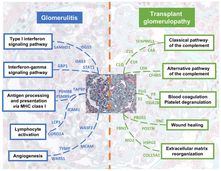
Antibody-mediated rejection (ABMR) is the leading cause of allograft failure in kidney transplantation. Its histological hallmark is represented by lesions of glomerulitis i.e., inflammatory cells within glomeruli. Current therapies for ABMR fail to prevent chronic allograft damage i.e., transplant glomerulopathy, leading to allograft loss. We used laser microdissection of glomeruli from formalin-fixed allograft biopsies combined with mass spectrometry-based proteomics to describe the proteome modification of 11 active and 10 chronic active ABMR cases compared to 8 stable graft controls. Of 1335 detected proteins, 77 were deregulated in glomerulitis compared to stable grafts, particularly involved in cellular stress mediated by interferons type I and II, leukocyte activation and microcirculation remodeling. Three proteins extracted from this protein profile, TYMP, WARS1 and GBP1, showed a consistent overexpression by immunohistochemistry in glomerular endothelial cells that may represent relevant markers of endothelial stress during active ABMR. In transplant glomerulopathy, 137 proteins were deregulated, which favor a complement-mediated mechanism, wound healing processes through coagulation activation and ultimately a remodeling of the glomerular extracellular matrix, as observed by light microscopy. This study brings novel information on glomerular proteomics of ABMR in kidney transplantation, and highlights potential targets of diagnostic and therapeutic interest.

Keywords: antibody-mediated rejection; glomerulus; kidney transplantation; proteomics; transplant glomerulopathy.


Conflict of interest statement

The authors of this manuscript have no conflict of interest to disclose.

Figures

Figure 1
Figure 1
Overall analytical strategy of the study. The histological hallmark of kidney active antibody-mediated rejection is glomerulitis i.e., inflammatory cells within the glomerular capillary loops. This lesion can be seen by light microscopy (yellow circle and arrow, Masson’s trichrome staining, original magnification ×400). Prolonged injuries lead to an expansion of the extracellular matrix (stained in green with Masson’s trichrome) and a duplication of the glomerular basement membrane called transplant glomerulopathy. These lesions are associated with graft dysfunction, proteinuria and ultimately allograft loss. Herein we performed a bottom-up proteomic approach on laser microdissected glomeruli during active ABMR, chronic active ABMR and stable graft patients from formalin-fixed and paraffin-embedded allograft biopsies. The key technical steps are presented by following the blue arrow. Abbreviations: ABMR, antibody-mediated rejection.
Figure 2
Figure 2
Characterization of the proteome of glomerulitis, with volcano plots showing significantly deregulated proteins in the two ABMR/SG comparisons (A) and a heatmap-like plot displaying the main enriched biological processes (B). (A) Protein abundances were compared using Mann-Whitney U tests with a Benjamini-Hochberg correction of the p-values. The significance threshold for adjusted p-values was set at 0.05. For clarity, only the deregulated proteins are showed in the volcano plots for each comparison. Overrepresentation was defined as a fold change above 1.5 and an underrepresentation when the fold change was lesser than 0.66. (B) Biological processes follow the Gene Ontology terminology, identified by performing overrepresentation analyzes using the enrichGO function of the clusterProfiler R package version 4.0.5. Only the biological processes involving overrepresented proteins of the aABMR/SG comparison are presented here. Fold changes (log2) are displayed with a color gradient. Number of proteins enriched in each biological process is presented as a bar plot on the right-hand side. Redundant biological processes were manually removed. Abbreviations: aABMR, active antibody-mediated rejection; caABMR, chronic active antibody-mediated rejection; SG, stable graft control.
Figure 3
Figure 3
Highlighting glomerulitis with illustrative cases of glomerular immunostains for WARS1, TYMP, GBP1, CORO1A and EFHD2 in active (left), chronic active antibody-mediated rejection (middle) and stable graft control cases (right), original magnification × 400 ((A)–(O), respectively). Corresponding proteomic results depending on groups are displayed on the right-hand side, with adjusted p-values for each comparison (Mann-Whitney U tests and Benjamini-Hochberg correction). (A,B) show a diffuse cytoplasmic overexpression of WARS1 on endothelial cells in a glomerulus of an active and chronic active ABMR cases, with a strong and moderate positivity, respectively, compared to the constitutive weak and segmental endothelial staining observed in the stable graft control (C). (D,E) show a diffuse and moderate to strong overexpression of TYMP in endothelial cells, but also a strong staining in endocapillary inflammatory cells (E). (F) Rare inflammatory cells are strongly stained for TYMP (arrow), without endothelial staining in the glomerulus. (G) shows a strong staining for GBP1 in endothelial (arrow) and inflammatory cells in the glomeruli (focus of glomerulitis at the top). (H) displays a segmental and weak to moderate staining in some endothelial (arrow) and inflammatory cells. (I) No specific staining for GBP1 in a stable graft case. (J,K) show a strong, nuclear and cytoplasmic staining for CORO1A in noticeably all inflammatory cells, wherever they are. Many positive cells are visible in the glomerulus of both ABMR cases, with a segmental and global lesion of glomerulitis, respectively. (L) shows the same expression pattern of CORO1A, but limited to a few inflammatory cells in the peritubular capillaries or the interstitium, and just one cell in the glomerulus (arrow). (M,N) show a moderate to strong, nuclear and cytoplasmic staining for EFHD2 antibody in some inflammatory cells in the glomeruli of both ABMR cases. Of note, stained inflammatory cells are also seen in the nearby peritubular capillaries. (O) shows a similar pattern of expression of EFHD2, but with globally much fewer positive cells, limited in the glomerulus to 2 moderately stained neutrophils (arrow). Abbreviations: ABMR, antibody-mediated rejection.
Figure 4
Figure 4
Characterization of the proteome of transplant glomerulopathy, with a heatmap-like plot displaying the main enriched biological processes (A) and a description of the extracellular matrix remodeling (B). (A) Biological processes follow the Gene Ontology terminology, identified by performing overrepresentation analyzes, using the enrichGO function of the clusterProfiler R package version 4.0.5. Only the biological processes involving overrepresented proteins of the caABMR/aABMR comparison are presented here. Fold changes (log2) are displayed with a color gradient. Number of proteins enriched in each biological process is presented as a bar plot on the right hand side. Redundant biological processes were manually removed. (B) Extracellular matrix proteins (ECM) differentially represented in transplant glomerulopathy. Fold changes are illustrated as a bar plot. A grey-scale gradient reflects the p-values. Each protein is annotated by its ECM category according to the Matrisome database. Abbreviations: aABMR, active antibody-mediated rejection; caABMR, chronic active antibody-mediated rejection; SG, stable graft control; ECM, extracellular matrix.
Figure 5
Figure 5
Integrative summary of the most relevant pathways enriched in glomerulitis and transplant glomerulopathy. Main upregulated proteins are depicted for each pathway. Please note that some proteins are shared between pathways, but we chose here to avoid any duplicate for clarity.

References

    1. Lamb K.E., Lodhi S., Meier-Kriesche H.-U. Long-term renal allograft survival in the United States: A critical reappraisal. Am. J. Transplant. 2011;11:450–462. doi: 10.1111/j.1600-6143.2010.03283.x. - DOI - PubMed
    1. Sellarés J., de Freitas D.G., Mengel M., Reeve J., Einecke G., Sis B., Hidalgo L.G., Famulski K., Matas A., Halloran P.F. Understanding the causes of kidney transplant failure: The dominant role of antibody-mediated rejection and nonadherence. Am. J. Transplant. 2012;12:388–399. doi: 10.1111/j.1600-6143.2011.03840.x. - DOI - PubMed
    1. Sis B., Halloran P.F. Endothelial transcripts uncover a previously unknown phenotype: c4d-negative antibody-mediated rejection. Curr. Opin. Organ Transplant. 2010;15:42–48. doi: 10.1097/MOT.0b013e3283352a50. - DOI - PubMed
    1. Sis B., Jhangri G.S., Bunnag S., Allanach K., Kaplan B., Halloran P.F. Endothelial Gene expression in kidney transplants with alloantibody indicates antibody-mediated damage despite lack of C4d staining. Am. J. Transplant. 2009;9:2312–2323. doi: 10.1111/j.1600-6143.2009.02761.x. - DOI - PubMed
    1. Loupy A., Haas M., Roufosse C., Naesens M., Adam B., Afrouzian M., Akalin E., Alachkar N., Bagnasco S., Becker J.U., et al. The banff 2019 kidney meeting report (I): Updates on and clarification of criteria for T cell- and antibody-mediated rejection. Am. J. Transplant. 2020;20:2318–2331. doi: 10.1111/ajt.15898. - DOI - PMC - PubMed