. 2022 Mar 3;10(3):589.
doi: 10.3390/biomedicines10030589.

The Interplay of Cx26, Cx32, Cx37, Cx40, Cx43, Cx45, and Panx1 in Inner-Ear Development of Yotari (dab1-/-) Mice and Humans

Affiliations

The Interplay of Cx26, Cx32, Cx37, Cx40, Cx43, Cx45, and Panx1 in Inner-Ear Development of Yotari (dab1-/-) Mice and Humans

Josip Lesko et al. Biomedicines. .

Abstract

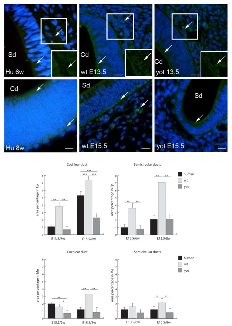
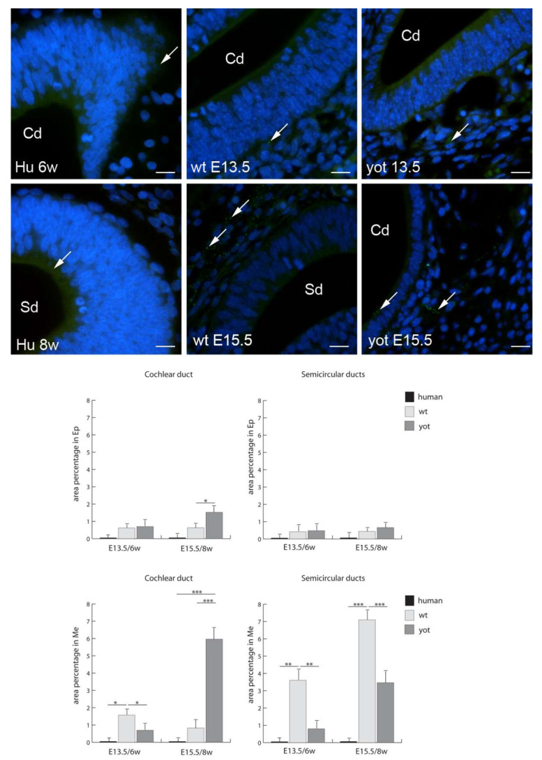
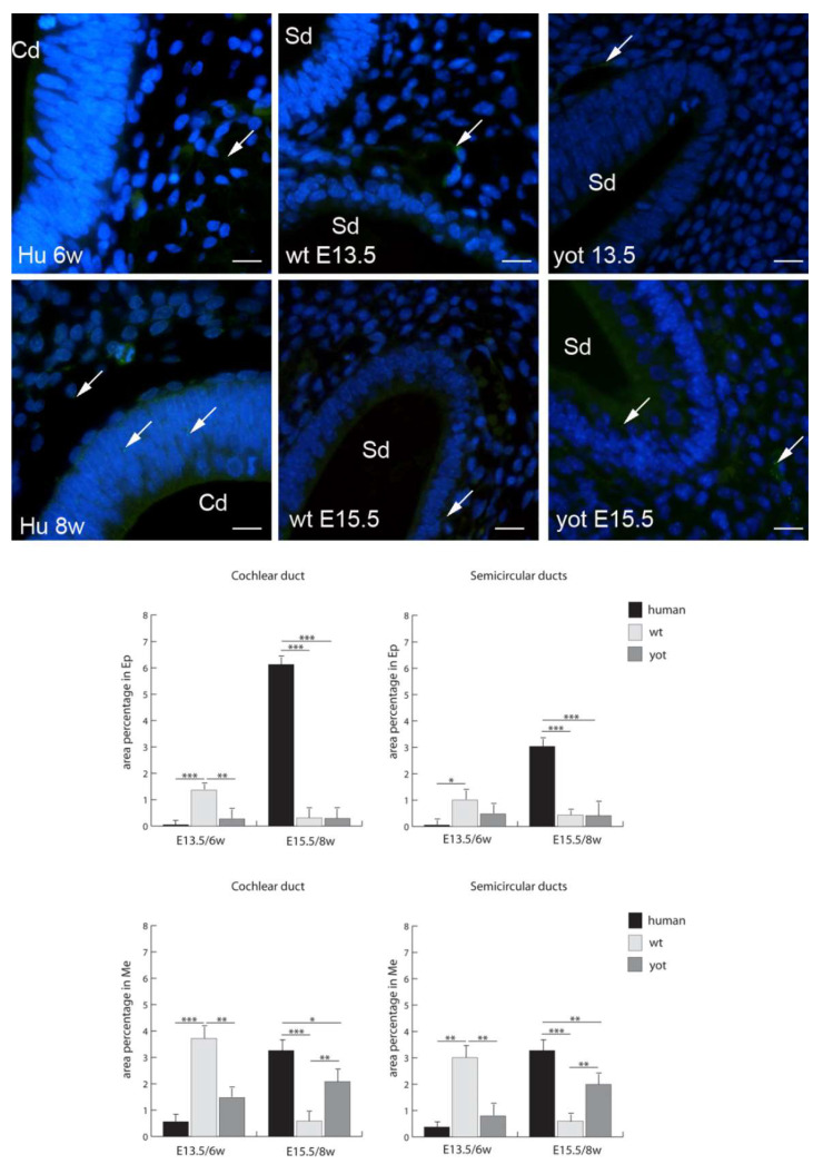
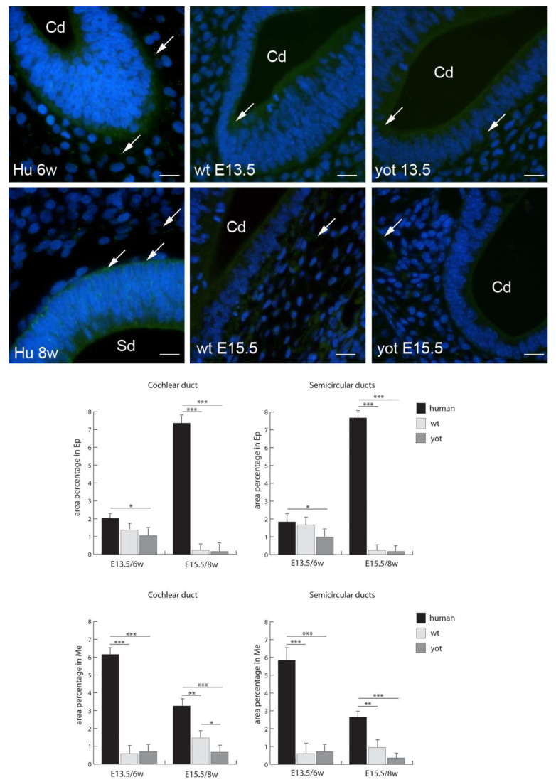
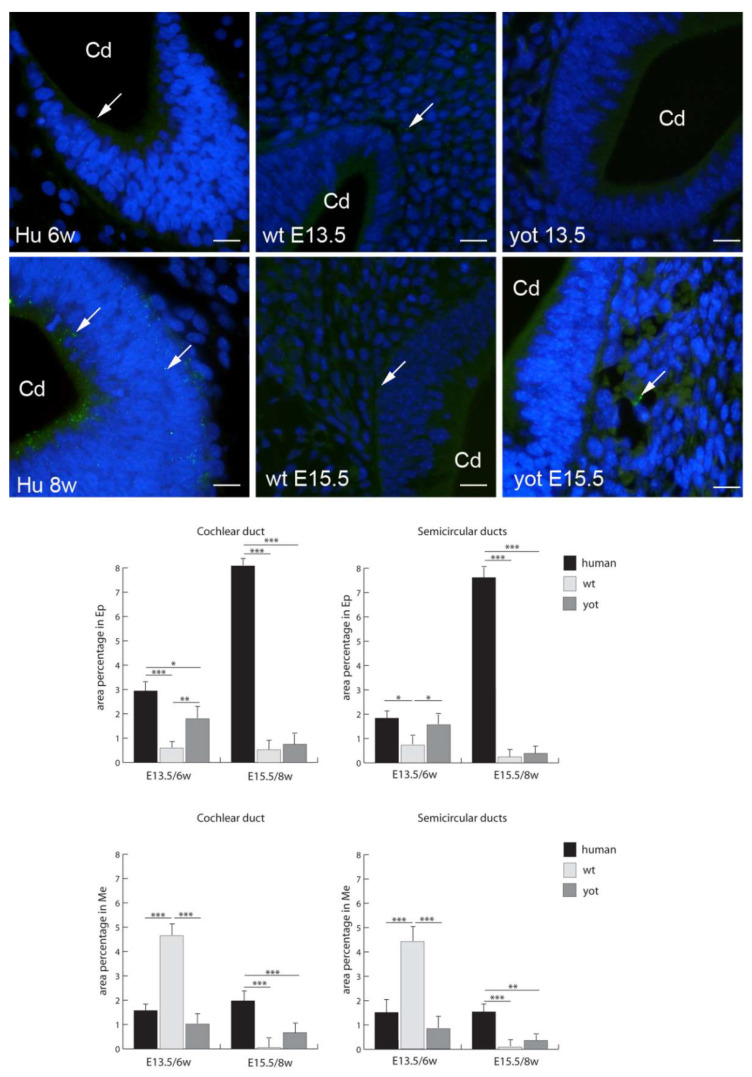
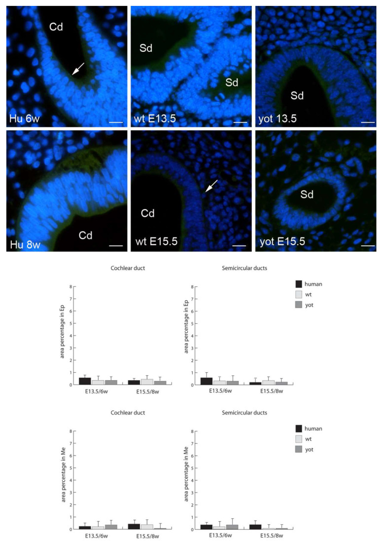
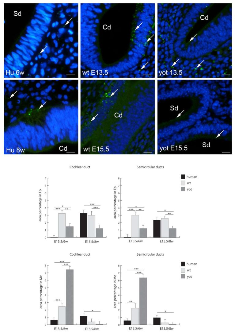
We investigated DAB1-protein deficiency in the inner-ear development of yotari in comparison to humans and wild-type (wt) mice by immunofluorescence for the expression of connexins (Cxs) and the pannexin Panx1. The spatial and temporal dynamics of Cx26, Cx32, Cx37, Cx40, Cx43, Cx45, and Panx1 were determined in the sixth and eighth weeks of human development and at the corresponding mouse embryonic E13.5 and E15.5, in order to examine gap junction intercellular communication (GJIC) and hemichannel formation. The quantification of the area percentage covered by positive signal was performed for the epithelium and mesenchyme of the cochlear and semicircular ducts and is expressed as the mean ± SD. The data were analysed by one-way ANOVA. Almost all of the examined Cxs were significantly decreased in the cochlear and semicircular ducts of yotari compared to wt and humans, except for Cx32, which was significantly higher in yotari. Cx40 dominated in human inner-ear development, while yotari and wt had decreased expression. The Panx1 expression in yotari was significantly lower than that in the wt and human inner ear, except at E13.5 in the mesenchyme of the wt and epithelium and mesenchyme of humans. Our results emphasize the relevance of GJIC during the development of vestibular and cochlear functions, where they can serve as potential therapeutic targets in inner-ear impairments.

Keywords: Cx26; Cx32; Cx37; Cx40; Cx43; Cx45; Panx1; gap junction; inner-ear development; yotari.


Conflict of interest statement

On behalf of all the authors, the corresponding author states that there is no conflict of interest.

Figures

Figure 1
Figure 1
Haematoxylin–eosin (HE) staining of the human, and wild-type (wt) and yotari mouse inner ears. HE staining of the human (first column), wt (second column), and yotari (third column) inner ears in the 6th (a,b) and 8th (c,d) weeks of development (6w/8w)/the embryonic day 13.5 (a,b) and E15.5 (c,d) (E13.5 and E15.5). Semicircular ducts (Sd), cochlear duct (Cd), saccule (s), utricle (u), vestibular ganglion (vg), spiral ganglion (sg), the cartilaginous shell (c), and the perilymphatic spaces (*). The scale bar for panels in the first (a) and third (c) rows equals 200 μm; that for panels in the second row (b) equals 40 μm, 100 μm, and 25 μm, respectively; that for panels in the fourth (d) row equals 100 μm.
Figure 2
Figure 2
Immunofluorescence staining of connexin Cx26 (arrows) in the human (Hu), and wild-type (wt) and yotari (yot) mouse inner ears in the 6th and 8th weeks of human development and at corresponding E13.5 and E15.5, merged with DAPI (blue nuclear stain). Scale bar is 10 µm, for all images. The panel with graphs represents the area percentages of connexin Cx26 in the human, and wild-type and yotari mouse inner ears in the 6th and 8th weeks of human development and at corresponding E13.5 and E15.5 in epithelium and mesenchyme of cochlear duct and semicircular ducts. Sd (semicircular ducts), Cd (cochlear duct), Ep (epithelium), and Me (mesenchyme). Data are presented as the mean ± SD (vertical line). Significant differences are indicated by * p < 0.05, ** p < 0.01, and *** p < 0.001. One-way ANOVA followed by Tukey’s multiple-comparisons test.
Figure 3
Figure 3
Immunofluorescence staining of connexin Cx32 (arrows) in the human (Hu), and wild-type (wt) and yotari (yot) mouse inner ears in the 6th and 8th weeks of human development and at corresponding E13.5 and E15.5, merged with DAPI (blue nuclear stain). Scale bar is 10 µm, for all images. The panel with graphs represents the area percentages of connexin Cx32 in the human, and wild-type and yotari mouse inner ears in the 6th and 8th weeks of human development and at corresponding E13.5 and E15.5 in epithelium and mesenchyme of cochlear duct and semicircular ducts. Sd (semicircular ducts), Cd (cochlear duct), Ep (epithelium), and Me (mesenchyme). Data are presented as the mean ± SD (vertical line). Significant differences are indicated by * p < 0.05, ** p < 0.01, and *** p < 0.001. One-way ANOVA followed by Tukey’s multiple-comparisons test.
Figure 4
Figure 4
Immunofluorescence staining of connexin Cx37 (arrows) in the human (Hu), and wild-type (wt) and yotari (yot) mouse inner ears in the 6th and 8th weeks of human development and at corresponding E13.5 and E15.5, merged with DAPI (blue nuclear stain). Scale bar is 10 µm, for all images. The panel with graphs represents the area percentages of connexin Cx37 in the human, and wild-type and yotari mouse inner ears at 6th and 8th weeks of human development and at corresponding E13.5 and E15.5 in epithelium and mesenchyme of cochlear duct and semicircular ducts. Sd (semicircular ducts), Cd (cochlear duct), Ep (epithelium), and Me (mesenchyme). Data are presented as the mean ± SD (vertical line). Significant differences are indicated by * p < 0.05, ** p < 0.01, and *** p < 0.001. One-way ANOVA followed by Tukey’s multiple-comparisons test.
Figure 5
Figure 5
Immunofluorescence staining of connexin Cx40 (arrows) in the human (Hu), and wild-type (wt) and yotari (yot) mouse inner ears in the 6th and 8th weeks of human development and at corresponding E13.5 and E15.5, merged with DAPI (blue nuclear stain). Scale bar is 10 µm, for all images. The panel with graphs represents the area percentages of connexin Cx40 in the human, and wild-type and yotari mouse inner ears in the 6th and 8th weeks of human development and at corresponding E13.5 and E15.5 in epithelium and mesenchyme of cochlear duct and semicircular ducts. Sd (semicircular ducts), Cd (cochlear duct), Ep (epithelium), and Me (mesenchyme). Data are presented as the mean ± SD (vertical line). Significant differences are indicated by * p < 0.05, ** p < 0.01, and *** p < 0.001. One-way ANOVA followed by Tukey’s multiple-comparisons test.
Figure 6
Figure 6
Immunofluorescence staining of connexin Cx43 (arrows) in the human (Hu), and wild-type (wt) and yotari (yot) mouse inner ears in the 6th and 8th weeks of human development and at corresponding E13.5 and E15.5, merged with DAPI (blue nuclear stain). Scale bar is 10 µm, for all images. The panel with graphs represents the area percentages of connexin Cx43 in the human, and wild-type and yotari mouse inner ears in the 6th and 8th weeks of human development and at corresponding E13.5 and E15.5 in epithelium and mesenchyme of cochlear duct and semicircular ducts. Sd (semicircular ducts), Cd (cochlear duct), Ep (epithelium), and Me (mesenchyme). Data are presented as the mean ± SD (vertical line). Significant differences are indicated by * p < 0.05, ** p < 0.01, and *** p < 0.001. One-way ANOVA followed by Tukey’s multiple-comparisons test.
Figure 7
Figure 7
Immunofluorescence staining of connexin Cx45 (arrows) in the human (Hu), and wild-type (wt) and yotari (yot) mouse inner ears in the 6th and 8th weeks of human development and at corresponding E13.5 and E15.5, merged with DAPI (blue nuclear stain). Scale bar is 10 µm, for all images. The panel with graphs represents the area percentages of connexin Cx45 in human, and wild-type and yotari mouse inner ears in the 6th and 8th weeks of human development and at corresponding E13.5 and E15.5 in epithelium and mesenchyme of cochlear duct and semicircular ducts. Sd (semicircular ducts), Cd (cochlear duct), Ep (epithelium), and Me (mesenchyme). Data are presented as the mean ± SD (vertical line). There were no significant differences according to one-way ANOVA and Tukey’s multiple-comparisons tests.
Figure 8
Figure 8
Immunofluorescence staining of pannexin 1 (Panx1) (arrows) in the human (Hu), and wild-type (wt) and yotari (yot) mouse inner ears in the 6th and 8th weeks of human development and at corresponding E13.5 and E15.5, merged with DAPI (blue nuclear stain). Scale bar is 10 µm, for all images. The panel with graphs represents the area percentages of Panx1 in the human, and wild-type and yotari mouse inner ears in the 6th and 8th weeks of human development and at corresponding E13.5 and E15.5 in epithelium and mesenchyme of cochlear duct and semicircular ducts. Sd (semicircular ducts), Cd (cochlear duct), Ep (epithelium), and Me (mesenchyme). Data are presented as the mean ± SD (vertical line). Significant differences are indicated by * p < 0.05, ** p < 0.01, and *** p < 0.001. One-way ANOVA followed by Tukey’s multiple-comparisons test.

Similar articles

Cited by

References

    1. Morsli H., Choo D., Ryan A., Johnson R., Wu D.K. Development of the mouse inner ear and origin of its sensory organs. J. Neurosci. Off. J. Soc. Neurosci. 1998;18:3327–3335. doi: 10.1523/JNEUROSCI.18-09-03327.1998. - DOI - PMC - PubMed
    1. Johnson Chacko L., Wertjanz D., Sergi C., Dudas J., Fischer N., Eberharter T., Hoermann R., Glueckert R., Fritsch H., Rask-Andersen H., et al. Growth and cellular patterning during fetal human inner ear development studied by a correlative imaging approach. BMC Dev. Biol. 2019;19:11. doi: 10.1186/s12861-019-0191-y. - DOI - PMC - PubMed
    1. Freyer L., Aggarwal V., Morrow B.E. Dual embryonic origin of the mammalian otic vesicle forming the inner ear. Development. 2011;138:5403–5414. doi: 10.1242/dev.069849. - DOI - PMC - PubMed
    1. Tafra R., Brakus S.M., Vukojevic K., Kablar B., Colovic Z., Saraga-Babic M. Interplay of proliferation and proapoptotic and antiapoptotic factors is revealed in the early human inner ear development. Otol. Neurotol. Off. Publ. Am. Otol. Soc. Am. Neurotol. Soc. Eur. Acad. Otol. Neurotol. 2014;35:695–703. doi: 10.1097/MAO.0000000000000210. - DOI - PubMed
    1. Falk M.M. Connexins/connexons. Cell-free expression. Methods Mol. Biol. 2001;154:91–116. - PMC - PubMed

LinkOut - more resources