Review
. 2022 Mar 3;10(3):602.
doi: 10.3390/biomedicines10030602.

Reactive Oxygen Species Induced Pathways in Heart Failure Pathogenesis and Potential Therapeutic Strategies

Affiliations
Review

Reactive Oxygen Species Induced Pathways in Heart Failure Pathogenesis and Potential Therapeutic Strategies

Aušra Mongirdienė et al. Biomedicines. .

Abstract

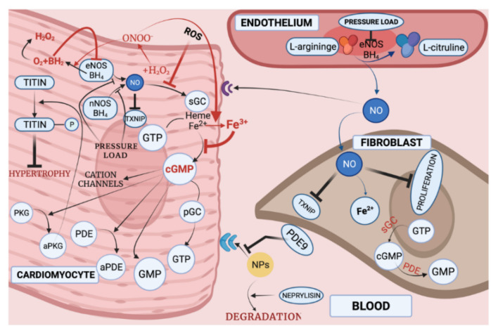
With respect to structural and functional cardiac disorders, heart failure (HF) is divided into HF with reduced ejection fraction (HFrEF) and HF with preserved ejection fraction (HFpEF). Oxidative stress contributes to the development of both HFrEF and HFpEF. Identification of a broad spectrum of reactive oxygen species (ROS)-induced pathways in preclinical models has provided new insights about the importance of ROS in HFrEF and HFpEF development. While current treatment strategies mostly concern neuroendocrine inhibition, recent data on ROS-induced metabolic pathways in cardiomyocytes may offer additional treatment strategies and targets for both of the HF forms. The purpose of this article is to summarize the results achieved in the fields of: (1) ROS importance in HFrEF and HFpEF pathophysiology, and (2) treatments for inhibiting ROS-induced pathways in HFrEF and HFpEF patients. ROS-producing pathways in cardiomyocytes, ROS-activated pathways in different HF forms, and treatment options to inhibit their action are also discussed.

Keywords: NO; cGC; heart failure with preserved ejection fraction; heart failure with reduced ejection fraction; protein kinases; reactive oxygen species.


Conflict of interest statement

The authors declare no conflict of interest.

Figures

Figure 1
Figure 1
Reactive oxygen species. O2•−-superoxide anion, OH-hydroxyl radical, H2O2-hydrogen peroxide, HOO-hydroperoxyle radical, H2O-water.
Figure 2
Figure 2
Harmful effects of ROS in cardiomyocytes (created with BioRender.com on 11 February 2022).
Figure 3
Figure 3
Enzymes involved in ROS production in cardiomyocytes and fibroblasts (created with BioRender.com on 7 February 2022). NOX2 is presented to be activated by endothelin and angiotensin II [58], by cytokines [59] and mechanical stress [60]. Increased NOX2 activation leads to cytoskeletal dysfunction in patients with CHF [61]. It was discovered that superoxide anions, produced by NOX, can oxidize and degrade hydrobiopterin-4 (BH4) leading to NOS uncoupling [62]. Nitric oxide synthase 3 (NOS3) uncoupling was observed in myocardium exposed to chronic pressure load. NOS3 catalyzes nitric oxide (NO) synthesis under physiological conditions. NO has an antihypertrophic effect. However, NOS3 is uncoupled with pressure load, and this, in turn, leads to reduction in tetrahydrobiopterin-4 concentration, increase in ROS production, and, as a consequence, to cardiomyocyte hypertrophy [63]. It was also shown that increase in ROS activates MAPK, leading to increased expression of proteins, such as ERK, JNK and P38, which are related to cardiomyocyte hypertrophy [64] (Figure 3). What is more, NOX-derived ROS may activate XOR [65]. Additionally, angiotensin II-induced signaling and isolated cardiomyocyte hypertrophy are dependent on NOX2 [66]. GTP-binding protein Rac-1 (involved in NOX activation), as described in the literature, is involved in isolated myocyte hypertrophy, induced by endothelin I, phenylephrine, angiotensin II [67] and norepinephrine [68]. AMPK—adenosine monophosphate activated protein kinase; AT1R—angiotensin II receptor; NOX-NADPH oxidase; BH4—dihydrobiopterin-4; NOS—nitric oxide synthase; NO—nitric oxide; MAPK—mitogen-activated protein kinase; XOR—xanthine oxidoreductase; AMP—adenosine monophosphate; GMP—guanosine monophosphate; Rac-1—GTP-binding protein; NHE-1—sodium/hydrogen exchanger-1; ERK—extracellular signal-regulated kinase; JNK-c—Jun N-terminal kinase; p38—a focal point of interactions of the serine/threonine kinases), MMP—matrix metalloproteinase.
Figure 4
Figure 4
ROS in HFrEF pathogenesis (created with BioRender.com on 9 February 2022). Decrease in O2 and nutrient supply in cardiomyocytes results in ROS overproduction. Increase in ROS leads to MMP activation and consequently fibroblast proliferation, vacuolar degeneration, excitation-contraction coupled protein oxidation (consequently leading to decrease in contractile function of cardiomyocytes) and oxidation of mitochondrial OMM and IMM proteins. OMM protein damage results in MAPK pathway activation, leading to apoptosis. IMM protein OPA1 oxidation (resulting in inhibition) with BNIP3 inhibition by ROS causes mitochondrial fragmentation. Both mitochondrial and other cytosol protein oxidation by ROS lead to decrease in ETC protein expression, resulting in ATP production decrease, resulting in poor contraction. Hyperacetylation of ETC complexes, fatty acid beta-oxidation and TCA cycle proteins lead to inhibition of its activity. SIRT, in healthy cardiomyocytes, inhibits hyperacetylation and activates AMPK-induced pathway, leading to enzyme-antioxidant synthesis which leads to hypertrophy inhibition. Excess of ROS inhibits beneficial SIRT effects. (EF—ejection fraction; MMP—matrix metalloproteinase; ROS—reactive oxygen species; BNIP3—mitochondrial mitophagy marker; OPA1—optic atrophy 1 protein; DRP1—dynamin-related protein 1; MFN2—mitofusin 2; MAPK—mitogen-activated protein kinase; ETC—electron transport chain; β-OX—beta-oxidation; SIRT—sirtuin family of NAD+-dependent deacetylases; TCA—tricarboxylic acid cycle; LKB1—liver kinase B1; AMP—adenosine monophosphate; AMPK—AMP activated protein kinase; SOD—superoxide dismutase; CAT—chloramphenicol acetyltransferase; UCP2—uncoupling protein 2).
Figure 5
Figure 5
NO–cGMP-PKG pathway in HFpEF development (created with BioRender.com on 9 February 2022). NO, produced in endothelium by eNOS in normal conditions, protects fibroblasts and cardiomyocytes from harmful proliferation. BH4 (hydrobiopterin-4) is required for eNOS action. NO acts via stimulation of cardiac sGC receptors (leading to cGMP synthesis). cGMP regulates phosphodiesterases (PDEs) and cGMP-dependent protein kinases (PKG). NO inhibits TXNIP, resulting in inhibition of apoptosis, however ROS inhibit this action. Oxidative stress shifts sGC towards an oxidized heme-free form which is unresponsive to endogenous and exogenous NO. Titin hypophosphorylation leads to hypertrophy of cardiomyocytes. Increased peroxynitrite concentrations, together with scarce NO availability, induce fibroblast proliferation. Due to uncoupled eNOS, superoxide production increases. In turn, low levels of NO react with superoxide to generate peroxynitrite. Peroxynitrite: (1) oxidizes BH4 to BH2 (BH2 inhibits eNOS), and (2) oxidizes Fe2+ to Fe3+ (Fe3+ inhibits cGMP production from GTP). Therefore, cGMP cannot activate PKG to phosphorylate titin, whereas titin phosphate prevents cardiomyocyte hypertrophy. For this reason, eNOS inhibition results in both fibroblast and cardiomyocyte proliferation. Neprylisin catalyzes NPs degradation, and PDE9 inhibits NPs. NPs acts through receptors in cardiomyocytes to modulate proliferation of cardiomyocytes. NO—nitric oxide, eNOS—endothelial nitric oxide synthase, BH4—dihydrobiopterin-4, NPs—natriuretic peptide, PKG—protein kinase G, a—activated, sGC—soluble guanylyl cyclase, pGC—particulate guanylyl cyclase, PDE-GMP—regulated phosphodiesterase, ROS—reactive oxygen species, titin-P—phosphorylated titin, GTP—guanosine triphosphate, TXNIP—thioredoxin-interacting protein.

Similar articles

Cited by

References

    1. Holland D., Kumbhani D.J., Ahmed S.H., Marwick T.H. Effects of Treatment on Exercise Tolerance, Cardiac Function, and Mortality in Heart Failure with Preserved Ejection Fraction: A Meta-Analysis. J. Am. Coll. Cardiol. 2011;57:1676–1686. doi: 10.1016/j.jacc.2010.10.057. - DOI - PubMed
    1. Henning R.J. Diagnosis and treatment of heart failure with preserved left ventricular ejection fraction. World J. Cardiol. 2020;12:7–25. doi: 10.4330/wjc.v12.i1.7. - DOI - PMC - PubMed
    1. Shah A.M., Mann D. In search of new therapeutic targets and strategies for heart failure: Recent advances in basic science. Lancet. 2011;378:704–712. doi: 10.1016/S0140-6736(11)60894-5. - DOI - PMC - PubMed
    1. Shah S., Kitzman D.W., Borlaug B., Van Heerebeek L., Zile M., Kass D.A., Paulus W.J. Phenotype-Specific Treatment of Heart Failure with Preserved Ejection Fraction. Circulation. 2016;134:73–90. doi: 10.1161/CIRCULATIONAHA.116.021884. - DOI - PMC - PubMed
    1. Ponikowski P., Voors A.A., Anker S.D., Bueno H., Cleland J.G.F., Coats A.J.S. 2016 ESC Guidelines for the diagnosis and treatment of acute and chronic heart failure: The Task Force for the diagnosis and treatment of acute and chronic heart failure of the European Society of Cardiology (ESC)Developed with the special contribution of. Eur. Heart J. 2016;37:2129–2200. doi: 10.1093/eurheartj/ehw128. - DOI - PubMed