

Proteomic Studies of Psoriasis
- PMID: 35327421
- PMCID: PMC8945259
- DOI: 10.3390/biomedicines10030619
Proteomic Studies of Psoriasis
Abstract
In this review paper, we discuss the contribution of proteomic studies to the discovery of disease-specific biomarkers to monitor the disease and evaluate available treatment options for psoriasis. Psoriasis is one of the most prevalent skin disorders driven by a Th17-specific immune response. Although potential patients have a genetic predisposition to psoriasis, the etiology of the disease remains unknown. During the last two decades, proteomics became deeply integrated with psoriatic research. The data obtained in proteomic studies facilitated the discovery of novel mechanisms and the verification of many experimental hypotheses of the disease pathogenesis. The detailed data analysis revealed multiple differentially expressed proteins and significant changes in proteome associated with the disease and drug efficacy. In this respect, there is a need for proteomic studies to characterize the role of the disease-specific biomarkers in the pathogenesis of psoriasis, develop clinical applications to choose the most efficient treatment options and monitor the therapeutic response.
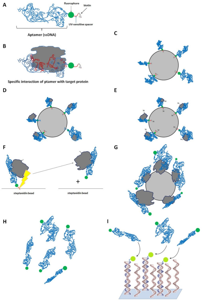
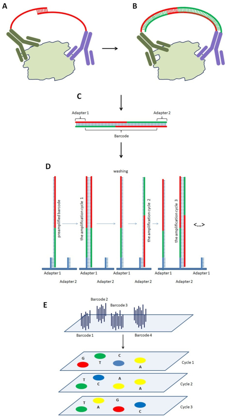
Keywords: LC-MS/MS; SOMAscan™; biomarkers; comorbidities; mass spectrometry; predisposition; proximity extension assay; psoriasis; risk factors.
Conflict of interest statement
The authors declare no conflict of interest.
Figures
Similar articles
-
Proteomics in Psoriasis.Int J Mol Sci. 2019 Mar 6;20(5):1141. doi: 10.3390/ijms20051141. Int J Mol Sci. 2019. PMID: 30845706 Free PMC article. Review.
-
Proteomic analysis of psoriatic skin lesions in a Chinese population.J Proteomics. 2021 May 30;240:104207. doi: 10.1016/j.jprot.2021.104207. Epub 2021 Mar 30. J Proteomics. 2021. PMID: 33798793
-
LC-MS/MS analysis of lesional and normally looking psoriatic skin reveals significant changes in protein metabolism and RNA processing.PLoS One. 2021 May 26;16(5):e0240956. doi: 10.1371/journal.pone.0240956. eCollection 2021. PLoS One. 2021. PMID: 34038424 Free PMC article.
-
Proteomic plasma profile of psoriatic patients.J Pharm Biomed Anal. 2018 Jun 5;155:185-193. doi: 10.1016/j.jpba.2018.03.068. Epub 2018 Apr 4. J Pharm Biomed Anal. 2018. PMID: 29635173
-
Analytical approaches to assess metabolic changes in psoriasis.J Pharm Biomed Anal. 2021 Oct 25;205:114359. doi: 10.1016/j.jpba.2021.114359. Epub 2021 Sep 2. J Pharm Biomed Anal. 2021. PMID: 34509137 Review.
Cited by
-
A Comprehensive Review of Protein Biomarkers for Invasive Lung Cancer.Curr Oncol. 2024 Aug 23;31(9):4818-4854. doi: 10.3390/curroncol31090360. Curr Oncol. 2024. PMID: 39329988 Free PMC article. Review.
-
Mass Spectrometry-Based Proteomics Analysis Unveils PTPRS Inhibits Proliferation and Inflammatory Response of Keratinocytes in Psoriasis.Inflammation. 2025 Feb;48(1):89-103. doi: 10.1007/s10753-024-02044-z. Epub 2024 May 13. Inflammation. 2025. PMID: 38739342
-
Assessment of Treatment-Relevant Immune Biomarkers in Psoriasis and Atopic Dermatitis: Toward Personalized Medicine in Dermatology.J Invest Dermatol. 2023 Aug;143(8):1412-1422. doi: 10.1016/j.jid.2023.04.005. Epub 2023 Jun 20. J Invest Dermatol. 2023. PMID: 37341663 Free PMC article. Review.
-
Balancing efficacy and hepatotoxicity: a comprehensive review of oral medications in psoriasis management.Naunyn Schmiedebergs Arch Pharmacol. 2025 Jun 25. doi: 10.1007/s00210-025-04334-1. Online ahead of print. Naunyn Schmiedebergs Arch Pharmacol. 2025. PMID: 40560394 Review.
-
The Expression of Cytokines and Chemokines Potentially Distinguishes Mild and Severe Psoriatic Non-Lesional and Resolved Skin from Healthy Skin and Indicates Different Stages of Inflammation.Int J Mol Sci. 2024 Oct 20;25(20):11292. doi: 10.3390/ijms252011292. Int J Mol Sci. 2024. PMID: 39457071 Free PMC article.
References
-
- Mogulevtseva J.A., Mezentsev A.V., Bruskin S.A. A Closer Look at Metalloproteinases. Nova Science Publishers; Hauppauge, NY, USA: 2019. The Role of Matrix Metalloproteinases in the Pathogenesis of Psoriasis; pp. 97–130.
Publication types
LinkOut - more resources
Full Text Sources
