

Disruption of the Ubiquitin-Proteasome System and Elevated Endoplasmic Reticulum Stress in Epilepsy
- PMID: 35327449
- PMCID: PMC8945847
- DOI: 10.3390/biomedicines10030647
Disruption of the Ubiquitin-Proteasome System and Elevated Endoplasmic Reticulum Stress in Epilepsy
Abstract
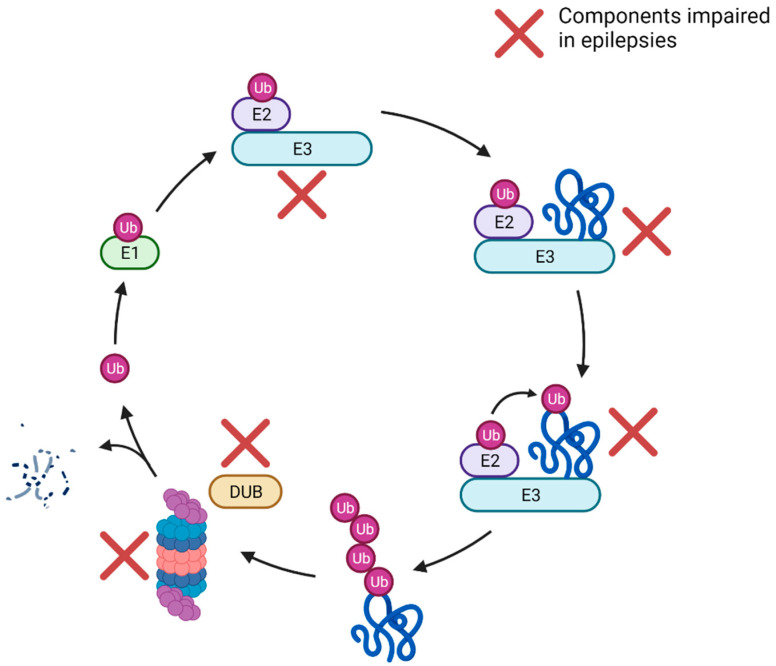
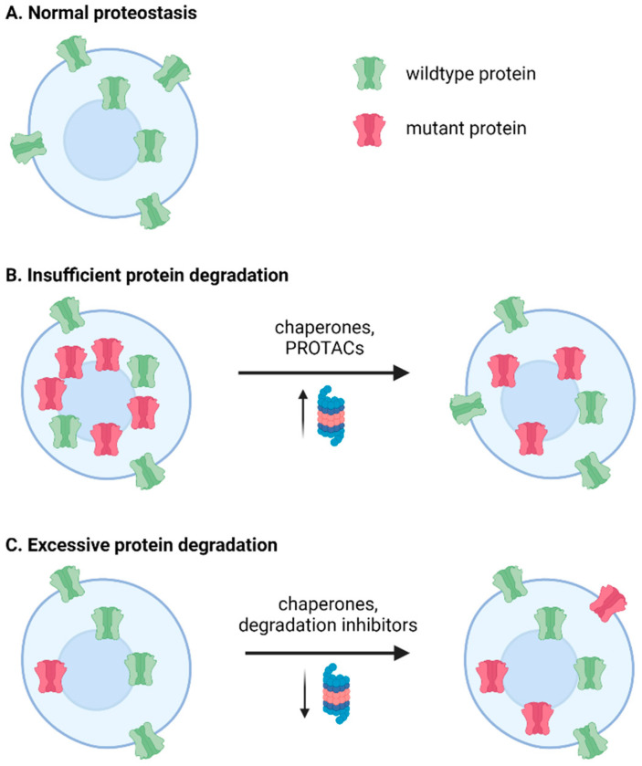
The epilepsies are a broad group of conditions characterized by repeated seizures, and together are one of the most common neurological disorders. Additionally, epilepsy is comorbid with many neurological disorders, including lysosomal storage diseases, syndromic intellectual disability, and autism spectrum disorder. Despite the prevalence, treatments are still unsatisfactory: approximately 30% of epileptic patients do not adequately respond to existing therapeutics, which primarily target ion channels. Therefore, new therapeutic approaches are needed. Disturbed proteostasis is an emerging mechanism in epilepsy, with profound effects on neuronal health and function. Proteostasis, the dynamic balance of protein synthesis and degradation, can be directly disrupted by epilepsy-associated mutations in various components of the ubiquitin-proteasome system (UPS), or impairments can be secondary to seizure activity or misfolded proteins. Endoplasmic reticulum (ER) stress can arise from failed proteostasis and result in neuronal death. In light of this, several treatment modalities that modify components of proteostasis have shown promise in the management of neurological disorders. These include chemical chaperones to assist proper folding of proteins, inhibitors of overly active protein degradation, and enhancers of endogenous proteolytic pathways, such as the UPS. This review summarizes recent work on the pathomechanisms of abnormal protein folding and degradation in epilepsy, as well as treatment developments targeting this area.
Keywords: ER stress; chaperones; epilepsy; proteostasis; ubiquitin proteasome system.
Conflict of interest statement
The authors declare no conflict of interest.
Figures
Similar articles
-
Perturbed proteostasis in autism spectrum disorders.J Neurochem. 2016 Dec;139(6):1081-1092. doi: 10.1111/jnc.13723. Epub 2016 Aug 4. J Neurochem. 2016. PMID: 27365114 Free PMC article. Review.
-
How Is the Fidelity of Proteins Ensured in Terms of Both Quality and Quantity at the Endoplasmic Reticulum? Mechanistic Insights into E3 Ubiquitin Ligases.Int J Mol Sci. 2021 Feb 19;22(4):2078. doi: 10.3390/ijms22042078. Int J Mol Sci. 2021. PMID: 33669844 Free PMC article. Review.
-
Targeting Ubiquitin-Proteasome Pathway by Natural Products: Novel Therapeutic Strategy for Treatment of Neurodegenerative Diseases.Front Physiol. 2020 Apr 28;11:361. doi: 10.3389/fphys.2020.00361. eCollection 2020. Front Physiol. 2020. PMID: 32411012 Free PMC article.
-
Alteration of the proteostasis network of plant cells promotes the post-endoplasmic reticulum trafficking of recombinant mutant (L444P) human β-glucocerebrosidase.Plant Signal Behav. 2014;9(3):e28714. doi: 10.4161/psb.28714. Epub 2014 Apr 8. Plant Signal Behav. 2014. PMID: 24713615 Free PMC article.
-
Adapting the endoplasmic reticulum proteostasis rescues epilepsy-associated NMDA receptor variants.Acta Pharmacol Sin. 2024 Feb;45(2):282-297. doi: 10.1038/s41401-023-01172-w. Epub 2023 Oct 6. Acta Pharmacol Sin. 2024. PMID: 37803141 Free PMC article.
Cited by
-
Pharmacological chaperones restore proteostasis of epilepsy-associated GABAA receptor variants.bioRxiv [Preprint]. 2023 Apr 19:2023.04.18.537383. doi: 10.1101/2023.04.18.537383. bioRxiv. 2023. Update in: Pharmacol Res. 2024 Oct;208:107356. doi: 10.1016/j.phrs.2024.107356. PMID: 37131660 Free PMC article. Updated. Preprint.
-
Role of inflammasomes and neuroinflammation in epilepsy.Immunol Rev. 2025 Jan;329(1):e13421. doi: 10.1111/imr.13421. Epub 2024 Nov 10. Immunol Rev. 2025. PMID: 39523682 Free PMC article. Review.
-
Pharmacological chaperones restore proteostasis of epilepsy-associated GABAA receptor variants.Pharmacol Res. 2024 Oct;208:107356. doi: 10.1016/j.phrs.2024.107356. Epub 2024 Aug 30. Pharmacol Res. 2024. PMID: 39216838 Free PMC article.
-
The "Cerebrospinal Fluid Sink Therapeutic Strategy" in Alzheimer's Disease-From Theory to Design of Applied Systems.Biomedicines. 2022 Jun 25;10(7):1509. doi: 10.3390/biomedicines10071509. Biomedicines. 2022. PMID: 35884814 Free PMC article. Review.
-
KCTD13-mediated ubiquitination and degradation of GluN1 regulates excitatory synaptic transmission and seizure susceptibility.Cell Death Differ. 2023 Jul;30(7):1726-1741. doi: 10.1038/s41418-023-01174-5. Epub 2023 May 4. Cell Death Differ. 2023. PMID: 37142655 Free PMC article.
References
-
- Di X.-J., Wang Y.-J., Han D.-Y., Fu Y.-L., Duerfeldt A.S., Blagg B.S.J., Mu T.-W. Grp94 Protein Delivers γ-Aminobutyric Acid Type A (GABAA) Receptors to Hrd1 Protein-Mediated Endoplasmic Reticulum-Associated Degradation. J. Biol. Chem. 2016;291:9526–9539. doi: 10.1074/jbc.M115.705004. - DOI - PMC - PubMed
Publication types
LinkOut - more resources
Full Text Sources
