

Preclinical Models of Brain Metastases in Breast Cancer
- PMID: 35327469
- PMCID: PMC8945440
- DOI: 10.3390/biomedicines10030667
Preclinical Models of Brain Metastases in Breast Cancer
Abstract
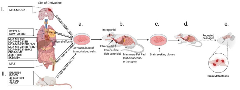
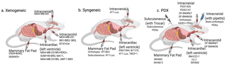
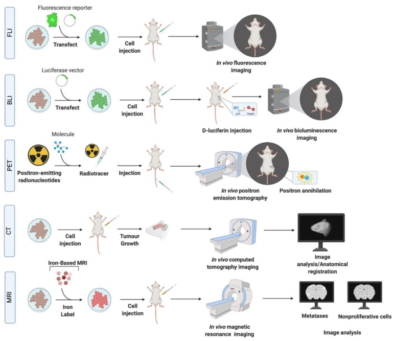
Breast cancer remains a leading cause of mortality among women worldwide. Brain metastases confer extremely poor prognosis due to a lack of understanding of their specific biology, unique physiologic and anatomic features of the brain, and limited treatment strategies. A major roadblock in advancing the treatment of breast cancer brain metastases (BCBM) is the scarcity of representative experimental preclinical models. Current models are predominantly based on the use of animal xenograft models with immortalized breast cancer cell lines that poorly capture the disease's heterogeneity. Recent years have witnessed the development of patient-derived in vitro and in vivo breast cancer culturing systems that more closely recapitulate the biology from individual patients. These advances led to the development of modern patient-tissue-based experimental models for BCBM. The success of preclinical models is also based on the imaging technologies used to detect metastases. Advances in animal brain imaging, including cellular MRI and multimodality imaging, allow sensitive and specific detection of brain metastases and monitoring treatment responses. These imaging technologies, together with novel translational breast cancer models based on patient-derived cancer tissues, represent a unique opportunity to advance our understanding of brain metastases biology and develop novel treatment approaches. This review discusses the state-of-the-art knowledge in preclinical models of this disease.
Keywords: animal imaging; brain metastasis; breast cancer; multimodal imaging; patient-derived xenografts; preclinical animal models.
Conflict of interest statement
The authors declare no conflict of interest.
Figures
Similar articles
-
Breast cancer brain metastasis: from etiology to state-of-the-art modeling.J Biol Eng. 2023 Jun 29;17(1):41. doi: 10.1186/s13036-023-00352-w. J Biol Eng. 2023. PMID: 37386445 Free PMC article. Review.
-
Improving orthotopic mouse models of patient-derived breast cancer brain metastases by a modified intracarotid injection method.Sci Rep. 2019 Jan 24;9(1):622. doi: 10.1038/s41598-018-36874-3. Sci Rep. 2019. PMID: 30679540 Free PMC article.
-
A common goal to CARE: Cancer Advocates, Researchers, and Clinicians Explore current treatments and clinical trials for breast cancer brain metastases.NPJ Breast Cancer. 2021 Sep 14;7(1):121. doi: 10.1038/s41523-021-00326-5. NPJ Breast Cancer. 2021. PMID: 34521857 Free PMC article. Review.
-
Patient-derived xenografts from non-small cell lung cancer brain metastases are valuable translational platforms for the development of personalized targeted therapy.Clin Cancer Res. 2015 Mar 1;21(5):1172-82. doi: 10.1158/1078-0432.CCR-14-1589. Epub 2014 Dec 30. Clin Cancer Res. 2015. PMID: 25549722
-
Comprehensive analysis of differentially expressed long noncoding RNAs, miRNAs and mRNAs in breast cancer brain metastasis.Epigenomics. 2021 Jul;13(14):1113-1128. doi: 10.2217/epi-2021-0152. Epub 2021 Jun 21. Epigenomics. 2021. PMID: 34148372
Cited by
-
Optimizing Precision Medicine for Breast Cancer Brain Metastases with Functional Drug Response Assessment.Cancer Res Commun. 2023 Jun 21;3(6):1093-1103. doi: 10.1158/2767-9764.CRC-22-0492. eCollection 2023 Jun. Cancer Res Commun. 2023. PMID: 37377606 Free PMC article.
-
Biological profile of breast cancer brain metastasis.Acta Neuropathol Commun. 2025 Apr 19;13(1):78. doi: 10.1186/s40478-025-01983-4. Acta Neuropathol Commun. 2025. PMID: 40253355 Free PMC article. Review.
-
State of the Art Modelling of the Breast Cancer Metastatic Microenvironment: Where Are We?J Mammary Gland Biol Neoplasia. 2024 Jul 16;29(1):14. doi: 10.1007/s10911-024-09567-z. J Mammary Gland Biol Neoplasia. 2024. PMID: 39012440 Free PMC article. Review.
-
Breast cancer brain metastasis: from etiology to state-of-the-art modeling.J Biol Eng. 2023 Jun 29;17(1):41. doi: 10.1186/s13036-023-00352-w. J Biol Eng. 2023. PMID: 37386445 Free PMC article. Review.
References
-
- Tevaarwerk A.J., Gray R.J., Schneider B.P., Smith M.L., Wagner L.I., Fetting J.H., Davidson N., Goldstein L.J., Miller K.D., Sparano J.A. Survival in patients with metastatic recurrent breast cancer after adjuvant chemotherapy: Little Evidence of Improvement over the Past 30 Years. Cancer. 2013;119:1140–1148. doi: 10.1002/cncr.27819. - DOI - PMC - PubMed
Publication types
LinkOut - more resources
Full Text Sources
