. 2022 Mar 15;10(3):676.
doi: 10.3390/biomedicines10030676.

Expression Profile of CD157 Reveals Functional Heterogeneity of Capillaries in Human Dermal Skin

Affiliations

Expression Profile of CD157 Reveals Functional Heterogeneity of Capillaries in Human Dermal Skin

Katarzyna Michalak-Micka et al. Biomedicines. .

Abstract

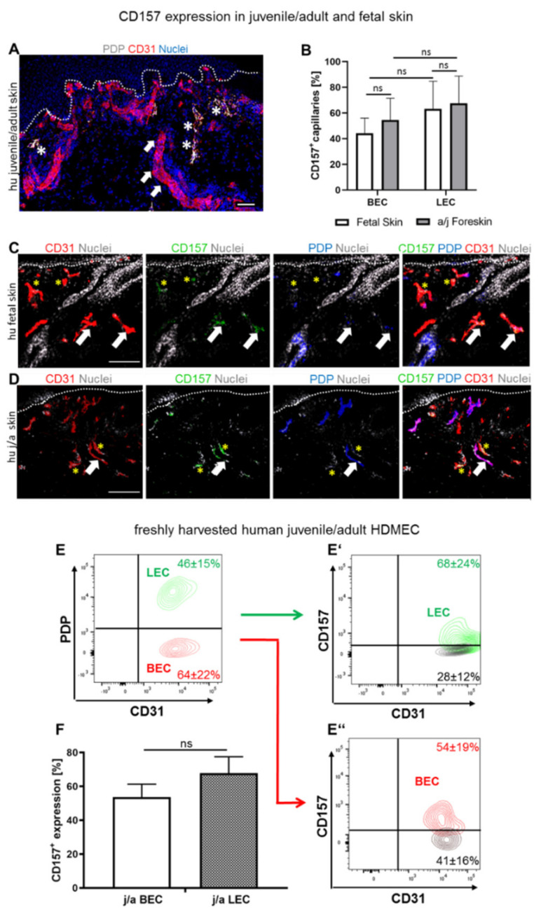
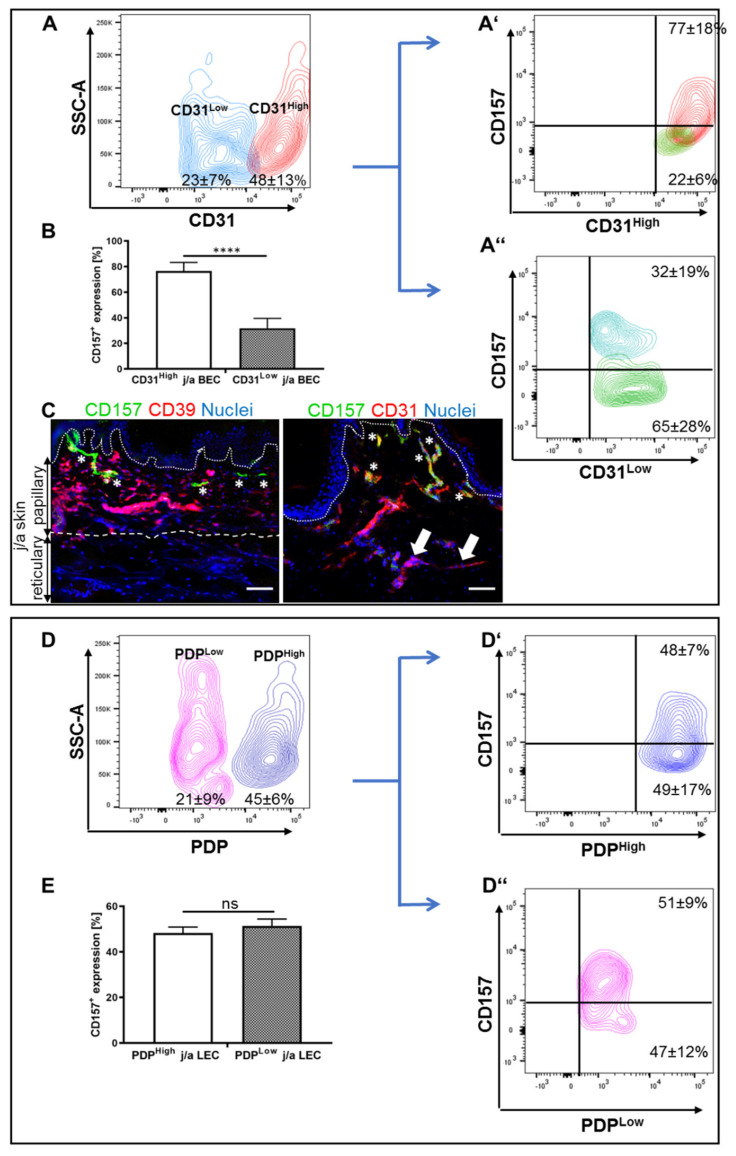
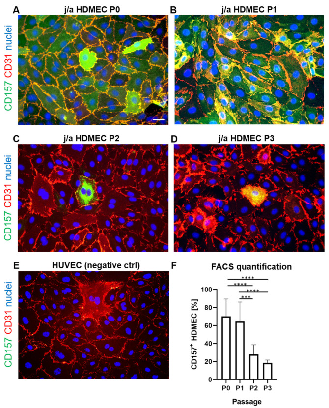
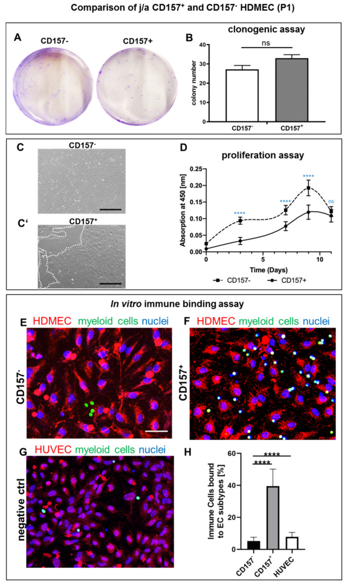
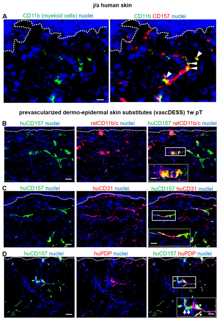
CD157 acts as a receptor, regulating leukocyte trafficking and the binding of extracellular matrix components. However, the expression pattern and the role of CD157 in human blood (BEC) and the lymphatic endothelial cells (LEC) of human dermal microvascular cells (HDMEC), remain elusive. We demonstrated constitutive expression of CD157 on BEC and LEC, in fetal and juvenile/adult skin, in situ, as well as in isolated HDMEC. Interestingly, CD157 epitopes were mostly localized on BEC, co-expressing high levels of CD31 (CD31High), as compared to CD31Low BEC, whereas the podoplanin expression level on LEC did not affect CD157. Cultured HDMEC exhibited significantly higher numbers of CD157-positive LEC, as compared to BEC. Interestingly, separated CD157- and CD157+ HDMEC demonstrated no significant differences in clonal expansion in vitro, but they showed distinct expression levels of cell adhesion molecules, before and after cytokine stimulation in vitro. In particular, we proved the enhanced and specific adherence of CD11b-expressing human blood myeloid cells to CD157+ HDMEC fraction, using an in vitro immune-binding assay. Indeed, CD157 was also involved in chemotaxis and adhesion of CD11b/c monocytes/neutrophils in prevascularized dermo-epidermal skin substitutes (vascDESS) in vivo. Thus, our data attribute specific roles to endothelial CD157, in the regulation of innate immunity during inflammation.

Keywords: CD157; CD157 receptor; angiogenesis; blood capillaries; immune cell adhesion; lymphatic capillaries; microvascular endothelial cells; myeloid cells; skin bio-engineering; vascular network formation.


Conflict of interest statement

The authors declare no conflict of interest.

Figures

Figure 1
Figure 1
CD157 expression in human juvenile/adult (j/a) and fetal (f) skin. (A) Representative immunofluorescence image used for the differentiation between blood (CD31+/PDPN; white arrows) and lymphatic (CD31+/PDPN+; white asterisks) capillaries (scale bar: 50 µm). Dotted line indocate dermo-epidermal junction. (B) The fraction of CD157+ capillaries in fetal (f) and juvenile/adult (j/a) foreskin skin sections was assessed based on the total number of blood and lymphatic capillaries (n = 7). (C,D) Human f and j/a skin was stained for endothelial-specific CD31, CD157 and PDPN markers, (scale bars: 100 µm). Asterisks indicate CD31+PDP-CD157+ capillaries, white arrows point to CD31+PDP+CD157+ capillaries. Dotted lines delineate dermo-epidermal junction. (EF) Sorting (E,E″) and quantification (F) of freshly isolated CD157+ and CD157BEC and LEC using FACS sorting (p = 0.6144, ns; n = 7). Statistical tests were performed using two-way ANOVA.
Figure 2
Figure 2
CD157 expression on CD31High/Low and podoplaninHigh/Low expressing freshly isolated HDMEC (A,B) The whole HDMEC population was separated into two distinct fractions: CD31High and CD31Low expressing subpopulations. (A′,A″,B) Representative FACS dot plots (A,A″) and quantification (B) of freshly isolated and separated cells. Whereas CD31High HDMEC showed enhanced expression of CD157+, CD31Low subpopulation showed a reduced expression of CD157 (**** p = 0.0010 extremely significant; n = 7). (C) Immunofluorescence co-staining for CD39, a papillary fibroblast marker (red) and CD157 (green) in j/a skin showing the presence of CD157 expressing capillaries (asterisks) in CD39-positive papillary dermis (C). Further, co-staining for CD31 (red) and CD157 (green) demonstrates CD157+CD31High capillaries (asterisks) in the upper, papillary dermis, whereas CD157-CD31Low capillaries (arrows) are predominantly located in lower, reticular dermal parts. (D,D″) HDMECs show distinct separation into PDPHigh and PDPLow expressing subpopulations. (E) Quantification of CD157 expression on PDPHigh and PDPLow HDMEC revealed no statistically significant differences between those two subpopulations (p = 0.530, ns; n = 7). Dotted lines delineate the dermo–epidermal junction. Dashed lines separate the papillary and reticular dermis. Statistical tests were performed using two-way ANOVA.
Figure 3
Figure 3
Expression profile of CD157 on cultured endothelial cells in vitro (AE) HDMEC cultured at passages 0 (A), 1 (B), 2 (C), and 3 (D) were stained for CD31 (red), CD157 (green), and counterstained for Hoechst (blue). HUVEC (E) were used as negative control. (F) A quantification using cell expression measured by flow cytometry, showed a similar high expression of CD157 with 69.2 ± 23.7% expressing cells at P0 and 64.5 ± 21.6% at P1; p = 0.96, ns). Further, strong decrease in CD157 expression levels was observed at P2 in vitro (27.9 ± 10.8% vs. P0; **** p < 0.0001 extremely significant), and at P3 (18.5 ± 3.2% vs. P0; **** p < 0.0001 extremely significant) (n = 10). *** p < 0.001 extremely significant. Statistical tests were performed using two-way ANOVA.
Figure 4
Figure 4
Comparison of clonogenic, proliferation, and immune binding potential of j/a CD157+ and CD157 HDMEC in vitro (A,B) CD157+ and CD157 HDMEC gave rise to similar colony numbers after sorting (p = 0.0589, ns; n = 5) (C,C′). Whereas CD157 HDMEC rapidly reached a confluent monolayer (C), CD157+ HDMECs demonstrated only a partial confluency (C′). White dashed lines delineate the areas not covered by cells (n = 5, scale bar: 50 µm). (D) Colorimetric proliferation assays with CD157+ and CD157 using 1 × 10⁴ seeded cells in 24-well plates were conducted at d = 0, 3, 7, 9, 11 (n = 5, p ≤ 0.0001). (EH) In vitro immune binding assay reveals strong adhesion of CD11b expressing myeloid cells to CD157+ (39.5 ± 10.7%), but only weak adhesion to CD157 (5.2 ± 2.4%, **** p < 0.0001 extremely significant) or HUVEC (7.8 ± 2.8%, **** p < 0.0001 extremely significant). Statistical tests were performed using two-way ANOVA.
Figure 5
Figure 5
Induction of HLA-DR and ICAM1 expression in CD157 and CD157+ BEC and LEC in vitro (AE) The protein expression of HLA-DR before and after 24-h (A), 48-h (B), and 72-h (C,E) stimulation of CD157 and CD157+ BEC and LEC with IFNγ and TNFα in vitro (n = 3). The expression level was assessed by flow cytomeric analysis (AC) and immunofluorescence staining (D,E). (FJ) The expression of ICAM1 before after 24-h (F), 48-h (G), and 72-h (H,J) stimulation of CD157 and CD157+ BEC and LEC with IFNγ and TNFα in vitro. The expression level was assessed by flow cytometric analysis (AC) and immunofluorescence staining (I,J) (n = 3). **** indicates p < 0.0001 extremely significant, *** indicates p < 0.001 extremely significant, ** indicates p < 0.01 very significant, * indicates p < 0.05 significant, ns indicates p ≥ 0.05 not significant. Statistical tests were performed using two-way ANOVA. Scale bars: 50 µm.
Figure 6
Figure 6
CD157 expression pattern on prevascularized dermo-epidermal skin substitutes (vascDESS) in vivo (A) Human myeloid CD11b-expressing cells in j/a foreskin sections co-localized with CD157+ capillaries (white arrowheads). (B) Multiple rat CD11b/c myeloid cells infiltrated vascDESS with the vast majority of them binding to CD157+ capillaries in dermal part in vivo after 1 week post-transplantation (pT). Insets represent magnified images of respective framed areas. (C,D) Immunofluorescence staining of in vivo transplants 1 week (w) post-transplantation (pT) confirm huCD157 expression on all human CD31 capillaries (n = 3 independent donors (C), including podoplanin-negative (PDP, white arrows) blood and podoplanin-positive (PDP+, white asterisks) lymphatic capillaries (n = 5). Scale bars: 50 µm (images) and 100 µm (inserts). Dotted lines delineate dermo-epidermal junction.

Similar articles

Cited by

References

    1. Lavagno L., Ferrero E., Ortolan E., Malavasi F., Funaro A. CD157 is part of a supramolecular complex with CD11b/CD18 on the human neutrophil cell surface. J. Biol. Regul. Homeost. Agents. 2007;21:5–11. doi: 10.3389/fimmu.2020.585294. - DOI - PubMed
    1. Ortolan E., Augeri S., Fissolo G., Musso I., Funaro A. CD157: From immunoregulatory protein to potential therapeutic target. Immunol. Lett. 2019;205:59–64. doi: 10.1016/j.imlet.2018.06.007. - DOI - PubMed
    1. Morone S., Augeri S., Cuccioloni M., Mozzicafreddo M., Angeletti M., Buono N.L., Giacomino A., Ortolan E., Funaro A. Binding of CD157 Protein to Fibronectin Regulates Cell Adhesion and Spreading. J. Biol. Chem. 2014;289:15588–15601. doi: 10.1074/jbc.M113.535070. - DOI - PMC - PubMed
    1. Ortolan E., Tibaldi E.V., Ferranti B., Lavagno L., Garbarino G., Notaro R., Luzzatto L., Malavasi F., Funaro A. CD157 plays a pivotal role in neutrophil transendothelial migration. Blood. 2006;108:4214–4222. doi: 10.1182/blood-2006-04-017160. - DOI - PubMed
    1. Wakabayashi T., Naito H., Suehiro J., Lin Y., Kawaji H., Iba T., Kouno T., Ishikawa-Kato S., Furuno M., Takara K., et al. CD157 Marks Tissue-Resident Endothelial Stem Cells with Homeostatic and Regenerative Properties. Cell Stem Cell. 2018;22:384–397. doi: 10.1016/j.stem.2018.01.010. - DOI - PubMed