

Putative Role of the Lung-Brain Axis in the Pathogenesis of COVID-19-Associated Respiratory Failure: A Systematic Review
- PMID: 35327531
- PMCID: PMC8944980
- DOI: 10.3390/biomedicines10030729
Putative Role of the Lung-Brain Axis in the Pathogenesis of COVID-19-Associated Respiratory Failure: A Systematic Review
Abstract
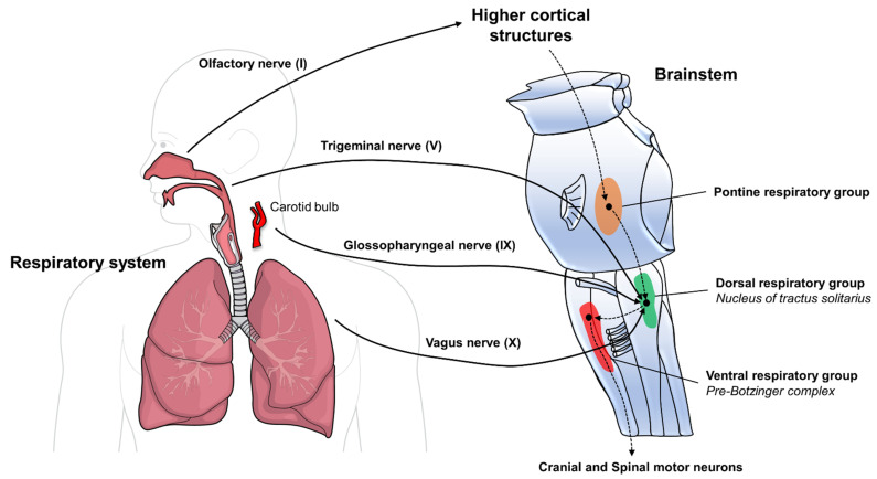
The emergence of SARS-CoV-2 and its related disease caused by coronavirus (COVID-19) has posed a huge threat to the global population, with millions of deaths and the creation of enormous social and healthcare pressure. Several studies have shown that besides respiratory illness, other organs may be damaged as well, including the heart, kidneys, and brain. Current evidence reports a high frequency of neurological manifestations in COVID-19, with significant prognostic implications. Importantly, emerging literature is showing that the virus may spread to the central nervous system through neuronal routes, hitting the brainstem and cardiorespiratory centers, potentially exacerbating the respiratory illness. In this systematic review, we searched public databases for all available evidence and discuss current clinical and pre-clinical data on the relationship between the lung and brain during COVID-19. Acknowledging the involvement of these primordial brain areas in the pathogenesis of the disease may fuel research on the topic and allow the development of new therapeutic strategies.
Keywords: COVID-19; SARS-CoV-2; acute respiratory distress syndrome; brainstem; neurological COVID; neuropathology; neurophysiology; respiratory failure; systematic review.
Conflict of interest statement
The authors declare no conflict of interest. The funders had no role in the design of the study; in the collection, analyses, or interpretation of data; in the writing of the manuscript, or in the decision to publish the results.
Figures
Similar articles
-
Computing the Effects of SARS-CoV-2 on Respiration Regulatory Mechanisms in COVID-19.ACS Chem Neurosci. 2020 Aug 19;11(16):2416-2421. doi: 10.1021/acschemneuro.0c00349. Epub 2020 Aug 11. ACS Chem Neurosci. 2020. PMID: 32600045 Free PMC article. Review.
-
Impact of Severe Acute Respiratory Syndrome Coronavirus 2 (SARS-CoV-2) in the Nervous System: Implications of COVID-19 in Neurodegeneration.Front Neurol. 2020 Nov 16;11:583459. doi: 10.3389/fneur.2020.583459. eCollection 2020. Front Neurol. 2020. PMID: 33304309 Free PMC article. Review.
-
Emerging COVID-19 Neurological Manifestations: Present Outlook and Potential Neurological Challenges in COVID-19 Pandemic.Mol Neurobiol. 2021 Sep;58(9):4694-4715. doi: 10.1007/s12035-021-02450-6. Epub 2021 Jun 24. Mol Neurobiol. 2021. PMID: 34169443 Free PMC article. Review.
-
Clinical manifestations and evidence of neurological involvement in 2019 novel coronavirus SARS-CoV-2: a systematic review and meta-analysis.J Neurol. 2020 Oct;267(10):2777-2789. doi: 10.1007/s00415-020-09974-2. Epub 2020 Jun 11. J Neurol. 2020. PMID: 32529575 Free PMC article.
-
Neuro-COVID-19: an insidious virus in action.Neurol Neurochir Pol. 2022;56(1):48-60. doi: 10.5603/PJNNS.a2021.0072. Epub 2021 Oct 13. Neurol Neurochir Pol. 2022. PMID: 34642927 Review.
Cited by
-
Database and AI Diagnostic Tools Improve Understanding of Lung Damage, Correlation of Pulmonary Disease and Brain Damage in COVID-19.Sensors (Basel). 2022 Aug 22;22(16):6312. doi: 10.3390/s22166312. Sensors (Basel). 2022. PMID: 36016071 Free PMC article.
-
Animal models of Long Covid: A hit-and-run disease.Sci Transl Med. 2024 Nov 13;16(773):eado2104. doi: 10.1126/scitranslmed.ado2104. Epub 2024 Nov 13. Sci Transl Med. 2024. PMID: 39536118 Free PMC article. Review.
-
Understanding the Pivotal Role of the Vagus Nerve in Health from Pandemics.Bioengineering (Basel). 2022 Jul 29;9(8):352. doi: 10.3390/bioengineering9080352. Bioengineering (Basel). 2022. PMID: 36004877 Free PMC article.
-
Long COVID in Children: A Multidisciplinary Review.Diagnostics (Basel). 2023 Jun 7;13(12):1990. doi: 10.3390/diagnostics13121990. Diagnostics (Basel). 2023. PMID: 37370884 Free PMC article. Review.
-
COVID-19 on Oral Health: A New Bilateral Connection for the Pandemic.Biomedicines. 2023 Dec 26;12(1):60. doi: 10.3390/biomedicines12010060. Biomedicines. 2023. PMID: 38255167 Free PMC article. Review.
References
Publication types
LinkOut - more resources
Full Text Sources
Miscellaneous
