Review
. 2022 Feb 28;12(3):382.
doi: 10.3390/biom12030382.

The Role of Membrane Lipids in Light-Activation of Drosophila TRP Channels

Affiliations
Review

The Role of Membrane Lipids in Light-Activation of Drosophila TRP Channels

Rita Gutorov et al. Biomolecules. .

Abstract

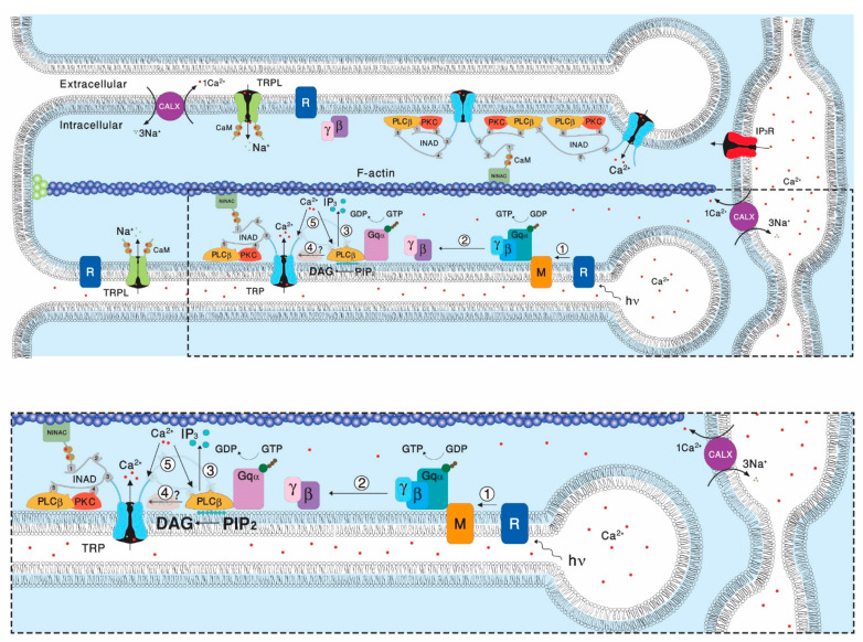
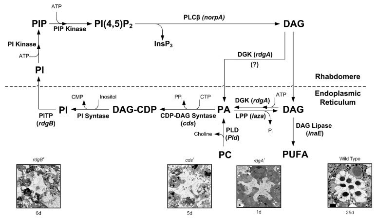
Transient Receptor Potential (TRP) channels constitute a large superfamily of polymodal channel proteins with diverse roles in many physiological and sensory systems that function both as ionotropic and metabotropic receptors. From the early days of TRP channel discovery, membrane lipids were suggested to play a fundamental role in channel activation and regulation. A prominent example is the Drosophila TRP and TRP-like (TRPL) channels, which are predominantly expressed in the visual system of Drosophila. Light activation of the TRP and TRPL channels, the founding members of the TRP channel superfamily, requires activation of phospholipase Cβ (PLC), which hydrolyzes phosphatidylinositol 4,5-bisphosphate (PIP2) into Diacylglycerol (DAG) and Inositol 1, 4,5-trisphosphate (IP3). However, the events required for channel gating downstream of PLC activation are still under debate and led to several hypotheses regarding the mechanisms by which lipids gate the channels. Despite many efforts, compelling evidence of the involvement of DAG accumulation, PIP2 depletion or IP3-mediated Ca2+ release in light activation of the TRP/TRPL channels are still lacking. Exogeneous application of poly unsaturated fatty acids (PUFAs), a product of DAG hydrolysis was demonstrated as an efficient way to activate the Drosophila TRP/TRPL channels. However, compelling evidence for the involvement of PUFAs in physiological light-activation of the TRP/TRPL channels is still lacking. Light-induced mechanical force generation was measured in photoreceptor cells prior to channel opening. This mechanical force depends on PLC activity, suggesting that the enzymatic activity of PLC converting PIP2 into DAG generates membrane tension, leading to mechanical gating of the channels. In this review, we will present the roles of membrane lipids in light activation of Drosophila TRP channels and present the many advantages of this model system in the exploration of TRP channel activation under physiological conditions.

Keywords: Diacylglycerol (DAG); Diacylglycerol kinase (DGK); Drosophila TRP channel; TRPL channel; cholesterol; ergosterol; methyl-β-cyclodextrin; phospholipase Cβ; poly unsaturated fatty acids (PUFAs).


Conflict of interest statement

Authors declare no conflict of interest.

Figures

Figure 1
Figure 1
The phosphoinositide cascade of vision. A diagram showing the molecular components of the signal transduction cascade of Drosophila: 1. Upon absorption of a photon (hν), rhodopsin (R) is converted into metarhodopsin (M). 2. The R to M photoconversion leads to the activation of heterotrimeric G protein (Gqαβγ) by promoting the GDP to GTP exchange. 3. The GTP-bound Gqα, in turn leads to activation of phospholipase Cβ (PLCβ), which hydrolyzes PIP2 into the soluble IP3 and the membrane bound DAG. 4. PLCβ in a still unclear way activates the TRP and TRPL channel, leading to an increase in microvillar Ca2+ concentration. 5. The increased Ca2+ concentration feeds back and negatively regulate both PLC and TRP channels activities. Elevation of DAG and Ca2+ promotes eye-specific protein kinase C (PKC) activity, which regulates channel activity. PLCβ, PKC, and the TRP ion channel form a supramolecular complex with the scaffolding protein INAD, which is bound to the F-actin cytoskeleton via the NINAC protein. Ca2+ level in the microvilli is also regulated by the Na+-Ca2+ exchanger, CALX. The diagram at the bottom is an amplification of the box marked by dotted lines in the upper diagram. (Modified from [14]).
Figure 2
Figure 2
The phosphoinositide cycle in Drosophila photoreceptors. In the phototransduction cascade, light triggers the activation of PLCβ. This catalyzes hydrolysis of the membrane PIP2 into water-soluble InsP3 and membrane-bound DAG. The continuous functionality of the photoreceptors during illumination is maintained by rapid regeneration of PIP2 in a cyclic enzymatic pathway (the PI cycle). DAG is transported by endocytosis to the endoplasmic reticulum (SMC) and inactivated by phosphorylation into PA via DGK. DGK may also be localized in the rhabdomeres. PA is converted to CDP–DAG via CDP-DAG syntase (encoded by the cds gene). PA can be converted back to DAG by lipid phosphate phosphohydrolase (LPP; also designated phosphatidic acid phosphatase (PAP) encoded by laza). PA is also produced from PC by PLD (encoded by Pld). DAG is hydrolyzed by DAG lipases, leading to the generation of PUFA. However, for PUFA generation, either an sn-2 DAG lipase or an additional enzyme (mono-acyl glycerol (MAG) lipase) would be required, but there is no evidence of either in Drosophila photoreceptors. Nevertheless, sn-1 type DAG lipase (encoded by inaE) was isolated. This DAG lipase was predominantly localized outside the rhabdomeres. The above difficulties put in question the participation of PUFA in channel activation in vivo. Subsequently, CDP-DAG is converted into phosphatidyl inositol (PI), which is transferred back to the microvillar membrane, by the PITP (encoded by the rdgB gene). Both RDGA and RDGB proteins have been immunolocalized to the SMC at the base of the rhabdomere. PIP and PIP2 are produced at the microvillar membrane by PI kinase and PIP kinase, respectively. Bottom: The ultrastructure of highly degenerated ommatidia induced by three mutations in the genes rdgB, cds, rdgA, which appear in the upper scheme: and wild type for a comparison. (Modified from [43]).
Figure 3
Figure 3
Depletion of cellular ATP activated the TRP channels: Membrane currents are usually elicited by light in the present of ATP and NAD in the pipette. Omission of these agents, combined with either few light pulses or with application of DNP to the bath induced constitutive activation of the light sensitive channels as monitored by a sustained noisy inward current that had the characteristics of the TRP or TRPL-dependent current. None of these currents were observed in the double null mutant trpl;trpP343. (A) The typical light induced current (LIC) of a wild type (WT) cell in response to orange lights (Schott OG 590, attenuated by 1 log unit) in the absence of ATP and NAD in the pipette. Note that after 3 light-pulses the TRP channels open spontaneously (right). The light monitor is indicated above the measured current traces. (B) Membrane currents were recorded 30 s after establishing the whole-cell configuration with physiological concentrations (1.5 mM) of Ca2+ in the bath. Whole cell recordings were conducted with pipettes, in which ATP and NAD were omitted and DNP was applied to the external medium at a time indicated by an arrow. Note the LIC was elicited by a light-pulse and the continuous opening of the channels in the dark following application of DNP. Also note that an additional light stimulus during the dark current elicited no response. (C): Families of current traces elicited by series of voltage steps from photoreceptors of wild type before (left traces) and following application of DNP (all other traces) in the presence of 1.5 mM Ca2+ in the bath (second family traces), with ~0 (nominal) mM Ca2+ in the bath (third family traces) and when 10 µM La3+ was applied to the bath (fourth family traces). For each experiment, a series of nine voltage steps was applied from a holding potential of −20 mV in 20 mV steps (bottom traces). (From [68], Copyright 2000 Society for Neuroscience).
Figure 4
Figure 4
Linoleic Acid (LA) removed Open Channel Block (OCB) from TRPL Channels: (A) Left, Representative I–V curves measured from S2 cells by whole cell patch clamp recordings using voltage ramps from −150 mV to 150 mV in 1s. The typical outward rectification of the TRPL channels (red curve 1) was modified to a linear I-V curve after application of 40 µM Linoleic Acid (LA, black curve 4). The effect at positive membrane potentials preceded that of negative membrane potentials (Curves 2 and 3, blue and purple respectively, see also 1A right). Application of 5 mM Ca2+ restored the outward rectifying I-V curve (green curve 5). Right: The current values at 90 mV (black dots) and −90 mV (dark red dots) are presented as a function of time. The numbers correspond to the curves presented in the left (n = 15). (B). Left, Representative IV curves measured as in A from mutant Drosophila ommatidia that express only TRPL channels (trpP343). In darkness the TRPL channels were closed (red curve 1). After application of 60 µM LA, a linear I-V curve was obtained (black curve 3). The effect of LA at positive membrane potentials preceded the effect at negative membrane potentials in a similar manner to expressed channels (blue curve 2). Application of 10 mM Ca2+ blocked the TRPL channel (green curve 4, left), ruling out the possibility that the linear I-V curve was due to leak current. Right, the effect of LA on the current is presented as a function of time at 90 mV (black dots) and −90 mV (dark red dots). The numbers correspond to the curves presented in the left (n = 5). The I-V curve of the LIC is presented in pink (the maximal light intensity was attenuated by 2 log units). (From [78], Copyright 2000 Society for Neuroscience)
Figure 5
Figure 5
The action of Linoleic Acid (LA) is not mediated via PLC: (A): PLC activity was monitored by using S2 cells expressing the Drosophila muscarinic receptor (DM1) and eGFP-PH, which binds to PIP2 and IP3. Application of 50µM LA, under conditions which activated the channels, did not elicit any change in the eGFP-PH distribution, as monitored by confocal images of the GFP fluorescence (LA) relative to control. Application of carbachol (CCH) elicited a robust translocation of eGFP-PH to the cytosol, which was reversed by CCH removal (wash), thus indicating activation of PLC (n = 6). The relative fluorescence intensity at a cross section of the cell (marked by line) is also presented below the confocal images. The time course of the fluorescence changes measured in the cytosol is presented on the right. (B). Western blot analysis of heads homogenate of dark raised WT and norpAp24;;trpP343 double null mutant flies. Head membrane was extracted with SDS buffer and subjected to Western blot analysis with antibodies specific for the Drosophila proteins TRP, NORPA and Dmoesin as indicated. No TRP and NORPA proteins were detected in the norpAp24;;trpP343 double mutant. (C). Left, Representative I-V curves measured by whole cell recordings from photoreceptors of the norpAP24;;trpP343 mutant lacking PLC and the TRP channel (see Figure 5B). In darkness the TRPL channels are closed (red curve 1). The effect of the 60µM LA was not altered by the absence of PLC and a linear I-V curve was obtained (black curve 4). The effect of LA at positive membrane potentials preceded that of negative membrane potentials (Curves 2 and 3, blue and purple respectively) in a similar manner to the results of S2. Application of 10 mM Ca2+ restored the outward rectifying I-V curve (green curve 5). Inset, enlargement of curves 1, 2 and 5, demonstrating more clearly the outward rectification. Right, the effect of LA on the current is presented at 90mV (black dots) and −90mV (dark red dots) as a function of time. The numbers correspond to the curves presented in the left (n = 4). (From [78], Copyright 2000 Society for Neuroscience).
Figure 6
Figure 6
Sequestration of cholesterol abolished the constitutive activity of TRPL channels. (A) Current–voltage relationships (I–V curves) measuring TRPL-dependent currents I–V curves obtained in response to voltage ramp (of 1 s duration) from S2 cells expressing TRPL and showing basal channel activity with strong outward rectification, typical for TRPL-dependent current (1). The TRPL channel activity was highly reduced after perfusion with 10 mM MβCD (2) and the effect was irreversible, even after washout of MβCD (3) (n > 10). (B) Time course of the MβCD effects on TRPL currents in S2 cells. Current densities are shown as a function of time. Series of I–V curves were derived from repeatedly applied voltage ramps every 5 s and currents were measured at ± 120 mV holding potentials as a function of time under the various experimental conditions as indicated. The numbers correspond to the numbers on the I–V curves in (A). (C) Statistics of the cholesterol depletion experiments in S2 cells. Cholesterol depletion by MβCD had a significant effect on the positive TRPL currents at 120 mV (n = 5, values are average ± SEM, paired Student t-test, * p ≤ 0.05). (From [121]).

Similar articles

Cited by

References

    1. Damann N., Voets T., Nilius B. TRPs in our senses. Curr. Biol. 2008;18:R880–R889. doi: 10.1016/j.cub.2008.07.063. - DOI - PubMed
    1. Hardie R.C., Raghu P. Visual transduction in Drosophila. Nature. 2001;413:186–193. doi: 10.1038/35093002. - DOI - PubMed
    1. Katz B., Minke B. Drosophila photoreceptors and signaling mechanisms. Front. Cell. Neurosci. 2009;3:2. doi: 10.3389/neuro.03.002.2009. - DOI - PMC - PubMed
    1. Kiselyov K., Patterson R.L. The integrative function of TRPC channels. Front. Biosci. 2009;14:45–58. doi: 10.2741/3230. - DOI - PubMed
    1. Minke B., Parnas M. Insights on TRP channels from in vivo studies in Drosophila. Annu. Rev. Physiol. 2006;68:649–684. doi: 10.1146/annurev.physiol.68.040204.100939. - DOI - PMC - PubMed

Publication types

MeSH terms

Substances

LinkOut - more resources