

Heat Stress Impairs Maternal Endometrial Integrity and Results in Embryo Implantation Failure by Regulating Transport-Related Gene Expression in Tongcheng Pigs
- PMID: 35327580
- PMCID: PMC8945854
- DOI: 10.3390/biom12030388
Heat Stress Impairs Maternal Endometrial Integrity and Results in Embryo Implantation Failure by Regulating Transport-Related Gene Expression in Tongcheng Pigs
Abstract
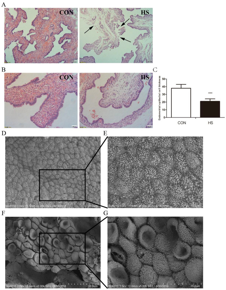
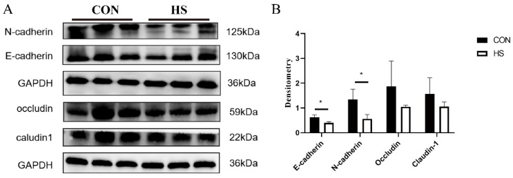
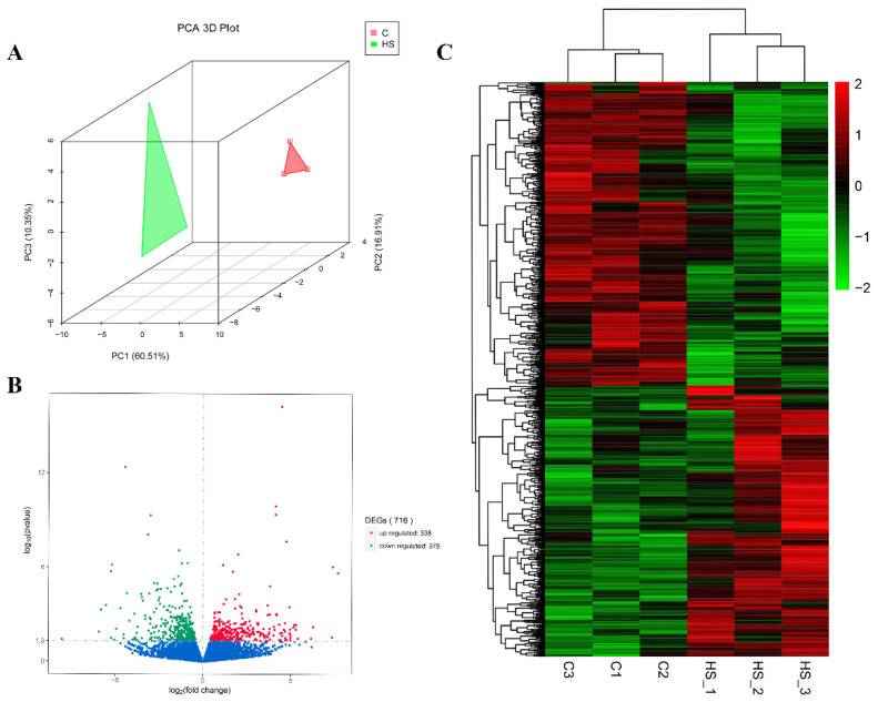
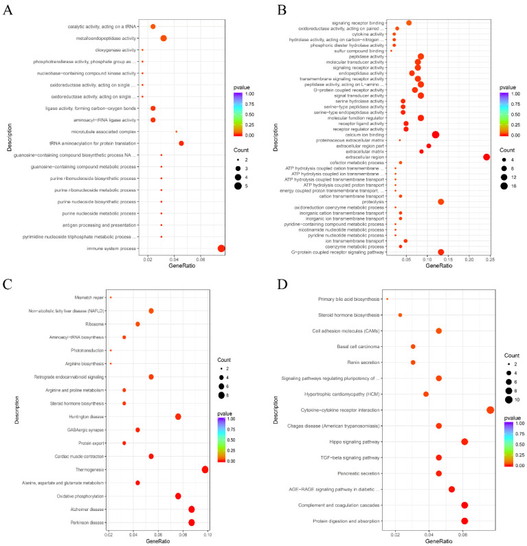
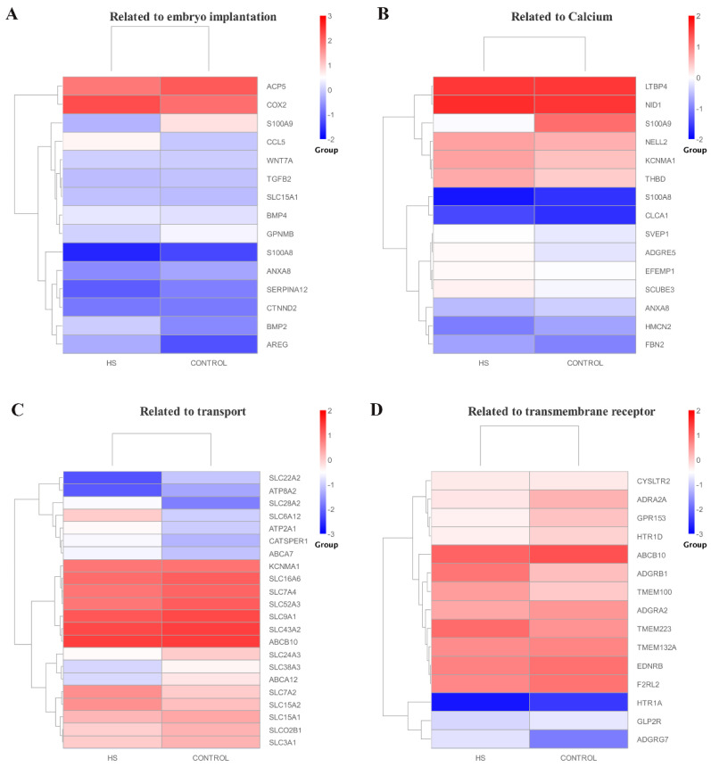
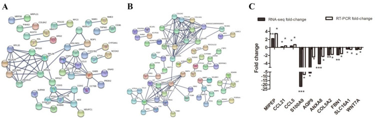
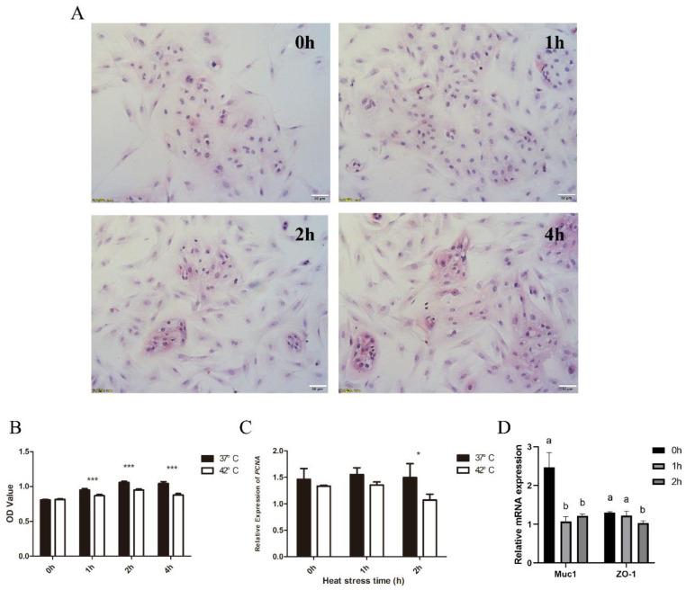
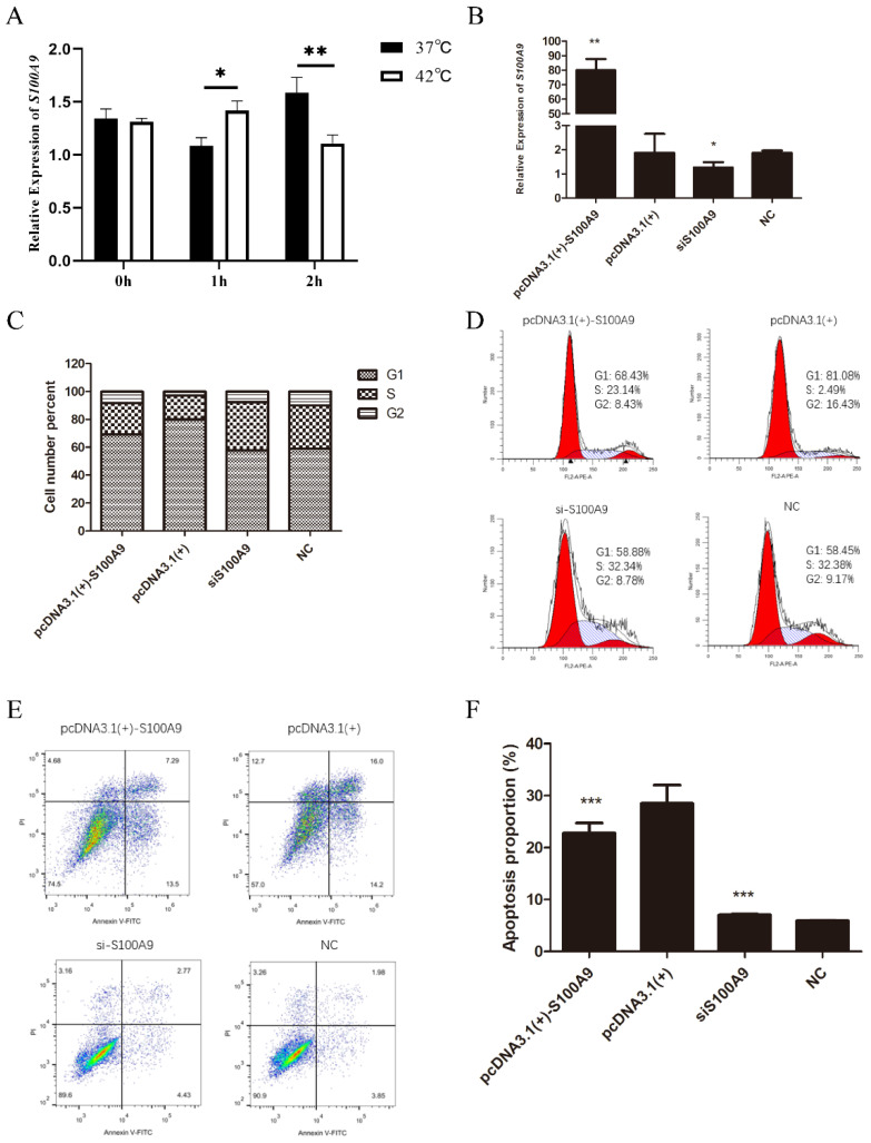
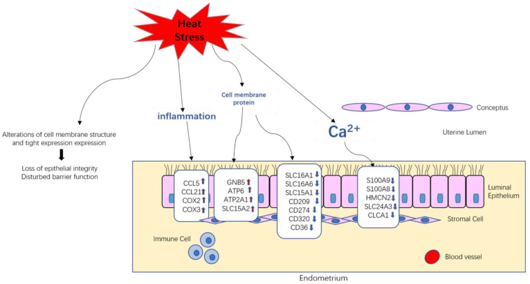
Heat stress (HS) poses a significant threat to production and survival in the global swine industry. However, the molecular regulatory effects of heat stress on maternal endometrial cells are poorly understood in pigs during early embryo implantation. In this study, we systematically examined morphological changes in the endometrium and the corresponding regulation mechanism in response to HS by combining scanning electron microscopy (SEM), hematoxylin/eosin (H&E) staining, western blot, and RNA-seq analyses. Our results showed that HS led to porcine endometrium damage and endometrial thinness during embryo implantation. The expression levels of cell adhesion-related proteins, including N-cadherin and E-cadherin, in the uterus were significantly lower in the heat stress group (39 ± 1 °C, n = 3) than in the control group (28 ± 1 °C, n = 3). A total of 338 up-regulated genes and 378 down-regulated genes were identified in porcine endometrium under HS. The down-regulated genes were found to be mainly enriched in the pathways related to the microtubule complex, immune system process, and metalloendopeptidase activity, whereas the up-regulated genes were mainly involved in calcium ion binding, the extracellular region, and molecular function regulation. S100A9 was found to be one of the most significant differentially expressed genes (DEGs) in the endometrium under HS, and this gene could promote proliferation of endometrial cells and inhibit their apoptosis. Meanwhile, HS caused endometrial epithelial cell (EEC) damage and inhibited its proliferation. Overall, our results demonstrated that HS induced uterine morphological change and tissue damage by regulating the expression of genes associated with calcium ions and amino acid transport. These findings may provide novel molecular insights into endometrial damage under HS during embryo implantation.
Keywords: S100A9; cell adhesion; heat stress; porcine endometrial cells; tight junction; transport activity.
Conflict of interest statement
The authors declare no conflict of interest.
Figures
Similar articles
-
Porcine endometrial heat shock proteins are differentially influenced by pregnancy status, heat stress, and altrenogest supplementation during the peri-implantation period.J Anim Sci. 2022 Jul 1;100(7):skac129. doi: 10.1093/jas/skac129. J Anim Sci. 2022. PMID: 35772767 Free PMC article.
-
Changes in calcium levels in the endometrium throughout pregnancy and the role of calcium on endometrial gene expression at the time of conceptus implantation in pigs.Mol Reprod Dev. 2019 Jul;86(7):883-895. doi: 10.1002/mrd.23166. Epub 2019 May 8. Mol Reprod Dev. 2019. PMID: 31066133
-
Spatial organization of endometrial gene expression at the onset of embryo attachment in pigs.BMC Genomics. 2019 Nov 21;20(1):895. doi: 10.1186/s12864-019-6264-2. BMC Genomics. 2019. PMID: 31752681 Free PMC article.
-
Ion channels in the endometrium: regulation of endometrial receptivity and embryo implantation.Hum Reprod Update. 2014 Jul-Aug;20(4):517-29. doi: 10.1093/humupd/dmu006. Epub 2014 Mar 2. Hum Reprod Update. 2014. PMID: 24591147 Review.
-
Embryo gene expression in pig pregnancy.Reprod Domest Anim. 2020 Apr;55(4):523-529. doi: 10.1111/rda.13647. Epub 2020 Feb 17. Reprod Domest Anim. 2020. PMID: 31986225 Review.
Cited by
-
Melatonin improves endometrial receptivity and embryo implantation via MT2/PI3K/LIF signaling pathway in sows.J Anim Sci Biotechnol. 2025 Jan 4;16(1):4. doi: 10.1186/s40104-024-01137-x. J Anim Sci Biotechnol. 2025. PMID: 39754262 Free PMC article.
-
Heat-Stress Impacts on Developing Bovine Oocytes: Unraveling Epigenetic Changes, Oxidative Stress, and Developmental Resilience.Int J Mol Sci. 2024 Apr 28;25(9):4808. doi: 10.3390/ijms25094808. Int J Mol Sci. 2024. PMID: 38732033 Free PMC article.
-
Enhancing uterine receptivity for embryo implantation through controlled collagenase intervention.Life Sci Alliance. 2024 Aug 16;7(10):e202402656. doi: 10.26508/lsa.202402656. Print 2024 Oct. Life Sci Alliance. 2024. PMID: 39151945 Free PMC article.
-
Effect of acute heat stress on intestinal immune response of Jinding ducks.Poult Sci. 2025 Aug;104(8):105273. doi: 10.1016/j.psj.2025.105273. Epub 2025 May 7. Poult Sci. 2025. PMID: 40367571 Free PMC article.
-
Functional transcriptome analysis revealed major changes in pathways affecting systems biology of Tharparkar cattle under seasonal heat stress.3 Biotech. 2024 Jul;14(7):177. doi: 10.1007/s13205-024-04018-2. Epub 2024 Jun 6. 3 Biotech. 2024. PMID: 38855148 Free PMC article.
References
-
- Lindenberg S.J. Experimental studies on the initial trophoblast endometrial interaction. Dan. Med. Bull. 1991;38:371–380. - PubMed
Publication types
MeSH terms
Substances
LinkOut - more resources
Full Text Sources
Research Materials
Miscellaneous
