. 2022 Mar 2;12(3):389.
doi: 10.3390/biom12030389.

Desmopressin Stimulates Nitric Oxide Production in Human Lung Microvascular Endothelial Cells

Affiliations

Desmopressin Stimulates Nitric Oxide Production in Human Lung Microvascular Endothelial Cells

Bianca Maria Rotoli et al. Biomolecules. .

Abstract

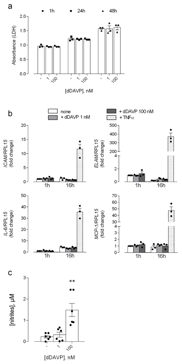
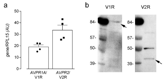
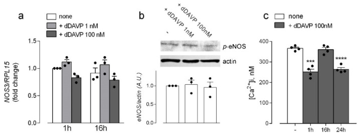
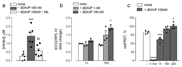
Desmopressin (dDAVP) is the best characterized analogue of vasopressin, the endocrine regulator of water balance endowed with potent vasoconstrictive effects. Despite the use of dDAVP in clinical practice, ranging from the treatment of nephrogenic diabetes insipidus to bleeding disorders, much remains to be understood about the impact of the drug on endothelial phenotype. The aim of this study was, thus, to evaluate the effects of desmopressin on the viability and function of human pulmonary microvascular endothelial cells (HLMVECs). The results obtained demonstrate that the vasopressor had no cytotoxic effect on the endothelium; similarly, no sign of endothelial activation was induced by dDAVP, indicated by the lack of effect on the expression of inflammatory cytokines and adhesion molecules. Conversely, the drug significantly stimulated the production of nitric oxide (NO) and the expression of the inducible isoform of nitric oxide synthase, NOS2/iNOS. Since the intracellular level of cAMP also increased, we can hypothesize that NO release is consequent to the activation of the vasopressin receptor 2 (V2R)/guanylate cyclase (Gs)/cAMP axis. Given the multifaceted role of NOS2-deriving NO for many physio-pathological conditions, the meanings of these findings in HLMVECs appears intriguing and deserves to be further addressed.

Keywords: NOS2; desmopressin; human endothelium; nitric oxide.


Conflict of interest statement

The authors declare no conflict of interest.

Figures

Figure 1
Figure 1
Effects of desmopressin on the viability and activation of microvascular endothelial cells. HLMVEC were incubated for the indicated times with different concentrations of dDAVP, as shown. (a). After the treatment, LDH release was assayed in the incubation medium as described in Methods. Data are the means ± SEM of four independent experiments each performed in triplicate. (b). The expression of the indicated genes was measured by means of RT-qPCR with the ∆∆Ct method and normalized for that of the housekeeping gene RPL15 (see Material and Methods). The stimulation with 10 ng/mL TNFα was employed for comparison, as positive control. (c). The production of NO was assessed through the quantitation of nitrites in the incubation medium, as described in Material and Methods. Data are means ± SEM of six independent determinations. ** p < 0.01 vs. control (-) with a two tailed Student’s t-test for unpaired data.
Figure 2
Figure 2
Expression of vasopressin receptors in microvascular endothelial cells. The expression of mRNA (a) and protein (b) for AVPR1A/V1R and AVPR2/V2R receptors was assessed in HLMVEC by means of RT-qPCR with the ∆∆Ct method and normalized for that of the housekeeping gene RPL15 (see Material and Methods). and Western Blot analysis, respectively, as described in Methods. Data in (a) are means ± SEM of four determinations, each performed in duplicate, while a representative blot is shown in (b) that, repeated three times, gave comparable results.
Figure 3
Figure 3
Arginine availability in desmopressin-treated human microvascular endothelial cells. HLMVEC were maintained for 24 h in the presence of the indicated concentrations of dDAVP. (a) Arginine intracellular concentration was measured through HPLC analysis after ninhydrin derivatization (see Material and Methods). Data are means ± SEM of three independent determinations. (b) Arginine uptake was assayed with a 30-s incubation of the cells in EBSS containing L-[3H]arginine (100 μM, 4 μCi/mL), as described in Material and Methods. Data are means ± SD of three determinations in a representative experiment that, repeated three times, gave comparable results.
Figure 4
Figure 4
Effects of desmopressin on the expression and activity of endothelial nitric oxide synthases in microvascular endothelial cells. HLMVEC were maintained in the presence of the indicated concentrations of dDAVP. (a) The expression of NOS3 was measured after 1 and 16 h with RT-qPCR and normalized for that of the housekeeping gene RPL15. Data are means ± SEM of three experiments, each performed in duplicate. (b) The phosphorylation of eNOS in Ser1177 was monitored by means of Western Blot analysis, as described in Material and Methods. A representative blot is shown that, repeated three times, gave comparable results; the graph represents the mean ± SEM of the results from the densitometric analyses of the different experiments (c) Intracellular calcium levels were measured in cells treated for the indicated times at steady-state and calibrated as described in Material and Methods. Data are mean ± SEM of four independent determinations. *** p < 0.001, **** p < 0.0001 vs. control (-), with a two tailed Student’s t-test for unpaired data.
Figure 5
Figure 5
Effects of desmopressin on the expression and activity of inducible nitric oxide synthases in microvascular endothelial cells. HLMVEC were maintained in the presence of the indicated concentrations of dDAVP. (a) The production of NO was assessed through the quantitation of nitrites in the incubation medium upon treatment for 48 h in the presence of iNOS inhibitor N6-(1-iminoethyl)-L-lysine (NIL; 100 μM). Data are means ± SEM of six independent determinations. (b) The expression of NOS2 was measured after 1 and 16 h with RT-qPCR and normalized for that of the housekeeping gene RPL15. Data are means ± SEM of three experiments, each performed in duplicate. (c) After the treatment, cells were transiently transfected with the EPAC-based FRET sensor target to cytosol (H96) or specifically to mitochondria (4mtH30) and the FRET analysis was performed as described (see Methods). Bars represent the mean ± SEM of four independent experiments. * p < 0.01, *** p < 0.001, **** p < 0.0001 vs. none, $$ p < 0.01 vs. dDAVP with a two tailed Student’s t-test for unpaired data.

Similar articles

Cited by

References

    1. Ball S.G. Vasopressin and disorders of water balance: The physiology and pathophysiology of vasopressin. Ann. Clin. Biochem. 2007;44:417–431. doi: 10.1258/000456307781646030. - DOI - PubMed
    1. Demiselle J., Fage N., Radermacher P., Asfar P. Vasopressin and its analogues in shock states: A review. Ann. Intensive Care. 2020;10:9. doi: 10.1186/s13613-020-0628-2. - DOI - PMC - PubMed
    1. Treschan T.A., Peters J. The vasopressin system: Physiology and clinical strategies. Anesthesiology. 2006;105:599–612. doi: 10.1097/00000542-200609000-00026. - DOI - PubMed
    1. Petersen M.B. The effect of vasopressin and related compounds at V1a and V2 receptors in animal models relevant to human disease. Basic Clin. Pharmacol. Toxicol. 2006;99:96–103. doi: 10.1111/j.1742-7843.2006.pto_299.x. - DOI - PubMed
    1. Manning M., Misicka A., Olma A., Bankowski K., Stoev S., Chini B., Durroux T., Mouillac B., Corbani M., Guillon G. Oxytocin and vasopressin agonists and antagonists as research tools and potential therapeutics. J. Neuroendocrinol. 2012;24:609–628. doi: 10.1111/j.1365-2826.2012.02303.x. - DOI - PMC - PubMed

Publication types

MeSH terms

LinkOut - more resources