Review
. 2022 Mar 4;12(3):404.
doi: 10.3390/biom12030404.

A Multisensory Network Drives Nuclear Mechanoadaptation

Affiliations
Review

A Multisensory Network Drives Nuclear Mechanoadaptation

Asier Echarri. Biomolecules. .

Abstract

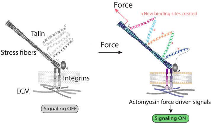
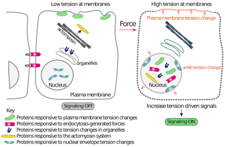
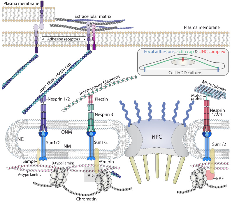
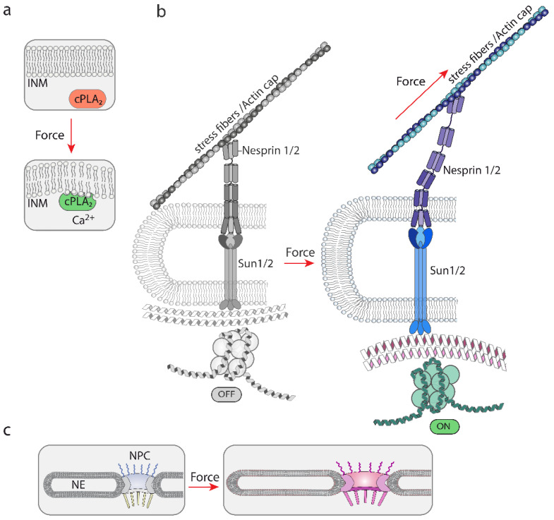
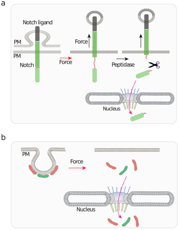
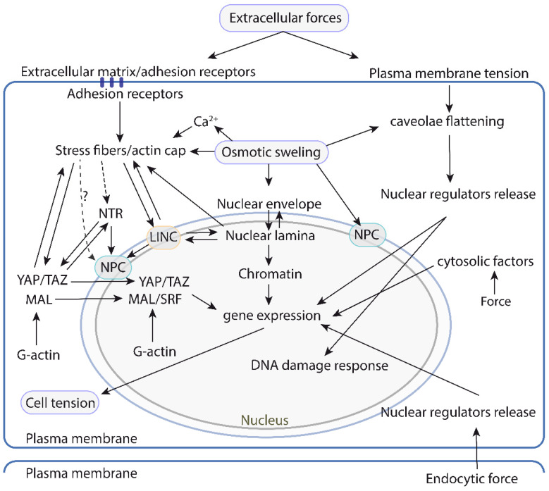
Cells have adapted to mechanical forces early in evolution and have developed multiple mechanisms ensuring sensing of, and adaptation to, the diversity of forces operating outside and within organisms. The nucleus must necessarily adapt to all types of mechanical signals, as its functions are essential for virtually all cell processes, many of which are tuned by mechanical cues. To sense forces, the nucleus is physically connected with the cytoskeleton, which senses and transmits forces generated outside and inside the cell. The nuclear LINC complex bridges the cytoskeleton and the nuclear lamina to transmit mechanical information up to the chromatin. This system creates a force-sensing macromolecular complex that, however, is not sufficient to regulate all nuclear mechanoadaptation processes. Within the nucleus, additional mechanosensitive structures, including the nuclear envelope and the nuclear pore complex, function to regulate nuclear mechanoadaptation. Similarly, extra nuclear mechanosensitive systems based on plasma membrane dynamics, mechanotransduce information to the nucleus. Thus, the nucleus has the intrinsic structural components needed to receive and interpret mechanical inputs, but also rely on extra nuclear mechano-sensors that activate nuclear regulators in response to force. Thus, a network of mechanosensitive cell structures ensures that the nucleus has a tunable response to mechanical cues.

Keywords: lipid bilayer mechano-sensing; mechano-transduction; mechanosensitive molecules; nuclear envelope; nucleus.


Conflict of interest statement

The author declares no conflict of interest.

Figures

Figure 1
Figure 1
A graphical representation of the ECM–integrin interface generated forces. Activation of integrins leads to mechanical unfolding of the actin cytoskeleton and integrin-bound talin. The current model suggests that activation of actin polymerization downstream of integrins exerts a pulling force on talin, unfolding its cryptic binding sites for vinculin, which starts a signaling cascade [13]. This represents one of the best understood examples of how mechanical forces change protein conformation, triggering biochemical modifications [9,13].
Figure 2
Figure 2
A graphical representation illustrating the different mechanisms acting independently on the ECM-integrin-actin-LINC-lamina-chromatin system that contributes to nuclear mechanoadaptation. Tension alterations in different membranes drive changes in the lipid bilayers that are sensed by proteins, which in turn modify their localization. This localization change drives enrichment in the nucleus or within nuclear regions, resulting in nuclear function regulation. Endocytic events also generate forces that induce downstream events leading to nuclear localization of the pathway effectors. The actomyosin system and forces reaching at organelles also regulate pathways that act independently of the LINC complex. These mechanisms are capable of transducing mechanical information to the nucleus by mechanisms that do not require nuclear-linked multimolecular structures, such as those shown in Figure 3 (ECM-integrin-actin cytoskeleton-LINC-lamina-chromatin).
Figure 3
Figure 3
A graphical representation of the main components of the chain of macromolecular complexes that transmit forces, initiated outside the cell, to the nucleus. The LINC complex, through nesprins, connects with the three cytoskeletons, which permits the nucleus to sense forces generated or sensed by these polymers. LINC: linker of nucleoskeleton and cytoskeleton. NPC: Nuclear pore complex. ONM: Outer nuclear membrane. INM: inner nuclear membrane. LAD: Lamin associated domains. The inset illustrates the actin cap bound to focal adhesions and the LINC complex while crossing over the nucleus.
Figure 4
Figure 4
A graphical representation of the different proposed mechanisms of mechanosensing by nuclear structures. (a) The NE is mechanosensitive. Tension changes modify the structure of the NE, which modifies the binding sites for the mechanosensitive cPLA2. Calcium accumulation contributes to the activation of cPLA2 [34]. Forces that alter lipid bilayers can be generated by osmolarity variations leading to changes in osmotic pressure. (b) A graphical representation of the model by which unfolding events in nesprin and SUN may lead to modifications in lamins and chromatin. This system is believed to rely on mechanical forces generated by actin polymerization and/or contraction of the actomyosin complex, which would pull from associated proteins, such as nesprins. These actions generate forces that would transmit mechanical cues up to the chromatin [2,40]. (c) A graphical representation illustrating the proposed sensitivity and adaptation of the NPC diameter to mechanical cues [41,42,43]. Hyperosmolarity [43] and ECM rigidity [42] have been shown to alter the NPC diameter.
Figure 5
Figure 5
A graphical representation of mechanosensitive events taking place at the plasma membrane with implications in nuclear functions. (a) Notch binding to its ligand initiates an endocytic process of the ligand, which exerts a pulling force on bound Notch. This stretching action unfolds Notch, unmasking cryptic peptidase sensitive sites, which triggers the release of a Notch fragment that is translocated to the nucleus to regulate gene expression [131]. (b) Caveolae are mechanosensitive plasma membrane (PM) invaginations that undergo flattening upon osmotic swelling [14,132,133]. This triggers the release, and subsequent nuclear translocation of EHD2 and Cavin3, represented as red and green modules, respectively, which regulate nuclear function [134].
Figure 6
Figure 6
A map of the physical and/or functional interactions between the different mechanosensitive or mechanoresponsive cell structures and pathways controlling nuclear mechanotransduction and function. The left side focuses on the pathways that rely on the actin cytoskeleton and the LINC complex to transduce mechanical information to the nucleus. This system is relatively well understood, and the current understanding suggests that it is highly interconnected with different mechanosensitive structures throughout. The right side illustrates the pathways that operate independently of the actin cytoskeleton-LINC axis and respond to forces generated or sensed at the plasma membrane. Mechanoresponsive events in cytosolic organelles that transduce to the nucleus have also been described and are depicted on the right side. LINC: linker of nucleoskeleton and cytoskeleton. NPC: Nuclear pore complex. NTR: nuclear transport receptors.

Similar articles

Cited by

References

    1. Vogel V., Sheetz M. Local force and geometry sensing regulate cell functions. Nat. Rev. Mol. Cell Biol. 2006;7:265–275. doi: 10.1038/nrm1890. - DOI - PubMed
    1. Kirby T.J., Lammerding J. Emerging views of the nucleus as a cellular mechanosensor. Nat. Cell Biol. 2018;20:373–381. doi: 10.1038/s41556-018-0038-y. - DOI - PMC - PubMed
    1. Iskratsch T., Wolfenson H., Sheetz M.P. Appreciating force and shape—The rise of mechanotransduction in cell biology. Nat. Rev. Mol. Cell Biol. 2014;15:825–833. doi: 10.1038/nrm3903. - DOI - PMC - PubMed
    1. Janota C.S., Calero-Cuenca F.J., Gomes E.R. The role of the cell nucleus in mechanotransduction. Curr. Opin. Cell Biol. 2020;63:204–211. doi: 10.1016/j.ceb.2020.03.001. - DOI - PubMed
    1. Moore S.W., Roca-Cusachs P., Sheetz M.P. Stretchy proteins on stretchy substrates: The important elements of integrin-mediated rigidity sensing. Dev. Cell. 2010;19:194–206. doi: 10.1016/j.devcel.2010.07.018. - DOI - PMC - PubMed

MeSH terms

LinkOut - more resources