. 2022 Mar 10;12(3):425.
doi: 10.3390/biom12030425.

Functional and Conformational Plasticity of an Animal Group 1 LEA Protein

Affiliations

Functional and Conformational Plasticity of an Animal Group 1 LEA Protein

Brett Janis et al. Biomolecules. .

Abstract

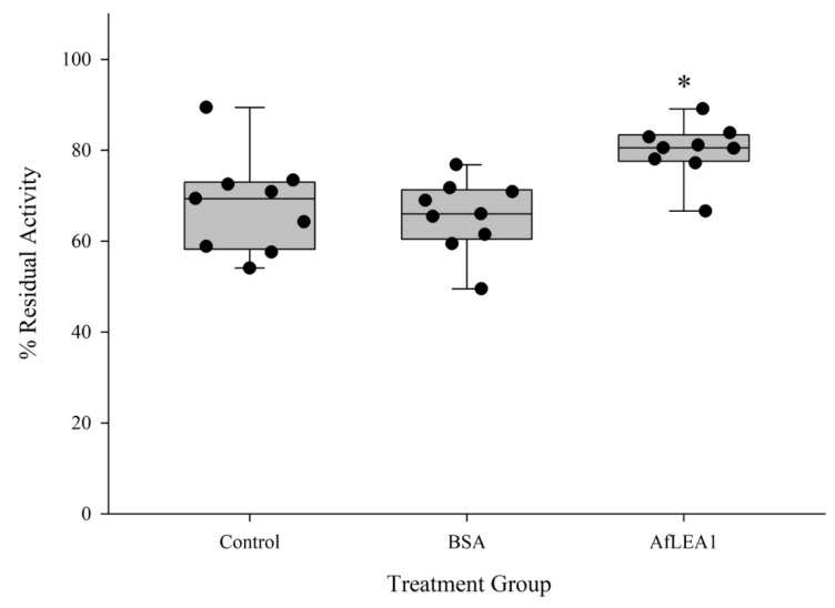
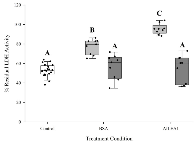
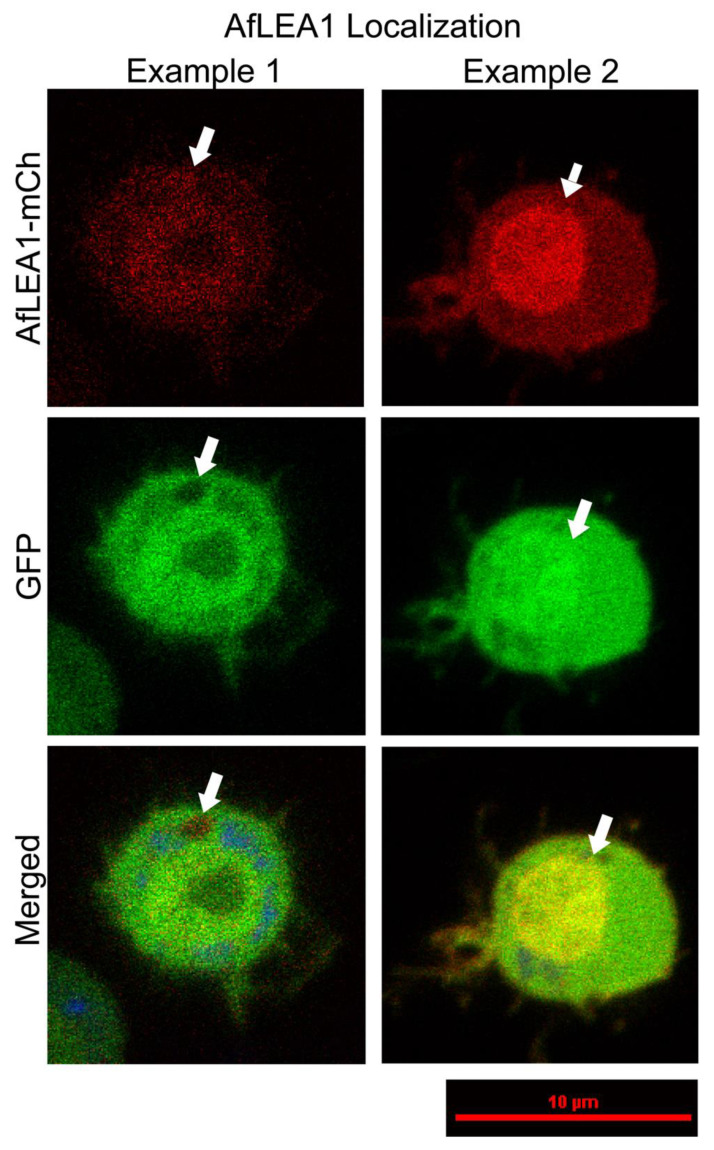
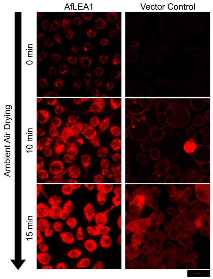
Group 1 (Dur-19, PF00477, LEA_5) Late Embryogenesis Abundant (LEA) proteins are present in organisms from all three domains of life, Archaea, Bacteria, and Eukarya. Surprisingly, Artemia is the only genus known to include animals that express group 1 LEA proteins in their desiccation-tolerant life-history stages. Bioinformatics analysis of circular dichroism data indicates that the group 1 LEA protein AfLEA1 is surprisingly ordered in the hydrated state and undergoes during desiccation one of the most pronounced disorder-to-order transitions described for LEA proteins from A. franciscana. The secondary structure in the hydrated state is dominated by random coils (42%) and β-sheets (35%) but converts to predominately α-helices (85%) when desiccated. Interestingly, AfLEA1 interacts with other proteins and nucleic acids, and RNA promotes liquid-liquid phase separation (LLPS) of the protein from the solvent during dehydration in vitro. Furthermore, AfLEA1 protects the enzyme lactate dehydrogenase (LDH) during desiccation but does not aid in restoring LDH activity after desiccation-induced inactivation. Ectopically expressed in D. melanogaster Kc167 cells, AfLEA1 localizes predominantly to the cytosol and increases the cytosolic viscosity during desiccation compared to untransfected control cells. Furthermore, the protein formed small biomolecular condensates in the cytoplasm of about 38% of Kc167 cells. These findings provide additional evidence for the hypothesis that the formation of biomolecular condensates to promote water stress tolerance during anhydrobiosis may be a shared feature across several groups of LEA proteins that display LLPS behaviors.

Keywords: LLPS; cryptobiosis; extremophiles; late embryogenesis abundant; protein condensate; water stress.


Conflict of interest statement

The authors declare that they have no conflict of interest.

Figures

Figure 1
Figure 1
Purification of AfLEA1. (A) SDS-PAGE of crude samples obtained after affinity chromatography (IMPACT) or further purification using anion-exchange chromatography as a polishing step. Fractions 1 and 2 obtained during anion-exchange chromatography contain AfLEA1 at the expected molecular weight of ~19 kDa. (B) AfLEA1 binds to quaternary ammonium groups at a pH of 8.0 and elutes in two distinct fractions (blue line) at relatively low concentrations of NaCl (black line).
Figure 2
Figure 2
Size-exclusion chromatography of AfLEA1, both by anion-exchange chromatography, obtained fractions of the AfLEA1 (blue: peak fraction 1; magenta: peak fraction 2) elute from a size-exclusion column between ovalbumin (44 kDa) and carbonic anhydrase (29 kDa), respectively. Based on these data, the apparent molecular mass of AfLEA1 computes to 33.4 kDa, or approximately 75% larger than the calculated mass based on the polypeptide sequence of the protein. The black line shows the elution of molecular weight markers. The elution maxima correspond to: (1) conalbumin, 75 kDa; (2) ovalbumin, 44 kDa; (3) carbonic anhydrase, 29 kDa; (4) ribonuclease A, 13.7 kDa; and (5) aprotinin 6.5 kDa.
Figure 3
Figure 3
Circular dichroism analysis of hydrated and desiccated AfLEA1, wherein α-helices (red), β-sheets (green), turns (blue), and random coils (magenta) are represented as proportions of the total protein structure. In the hydrated state, the secondary structure of AfLEA1 was on average 5% α-helices, 35% β-sheets, 18% turns, and 42% random coils. In the desiccated state, the secondary structure of AfLEA1 was on average 85% α-helices, 5% β-sheets, and 10% turns. In the presence of 2% SDS, the secondary structure of AfLEA1 was on average of 25% α-helices, 13% β-sheets, 16% turns, and 46% random coils (for CD spectra see Figure S2, Supplementary Materials).
Figure 4
Figure 4
Amino acid sequence features of AfLEA1 where the amino acid properties are labeled by colors representing aromatic (green), polar (yellow), nonpolar (grey), positive (blue), and negative (red) amino acid side chains. (A) The amino acid sequence of AfLEA1 is highly repetitive and consists of eight Group 1 LEA domains. (B) Projected as an α-helix, AfLEA1 has a distinct hydrophobic face of nonpolar amino acids (gray) but does not have distinctly organized stripes of charged amino acid as described for some Group 3 LEA proteins.
Figure 5
Figure 5
I-Tasser prediction of AfLEA1 structure in the desiccated state with amino acid properties labeled by color as polar (yellow), nonpolar (gray), aromatic (green), positive (red), and negative (blue). (A) AfLEA1 is predicted to fold into 84% α-helix, 5% β-sheet, and 11% turns. The tertiary structure of AfLEA1 is predicted to resemble a synthetic helical repeat protein (RCSB PDB ID: 5CWH) composed of helix-turn-helix structures, where each helix-turn-helix is a single Group 1 LEA motif. (B) The distribution of charged amino acids stabilizes the exterior of the AfLEA1. The protein’s interior is enriched with positive and aromatic residues.
Figure 6
Figure 6
Three-dimensional structure and charge distribution of three LEA proteins from A. franciscana as predicted by I-Tasser. Colors represent the positive-charge residues in red and negative residues in blue. (A) Predicted α-helical regions of AfrLEA2 are shown to have characteristic positive-negative-positive residue stripes c.f. [38]. (B) Predicted α-helical regions of AfrLEA3m also showed characteristic positive-negative-positive residue stripes c.f. [38]. (C) AfLEA1 does not present the proposed charge pattern observed in AfrLEA2 and AfrLEA3m in its secondary structure. Still, its tertiary structure presents adjacent stripes of an alternating formal charge (positive-negative-positive-negative) on the protein surface.
Figure 7
Figure 7
AfLEA1, dissolved in ultrapure water, crystallized readily when slowly desiccated at 80% RH. (A) Light microscopy shows that AfLEA1 dries into branching crystals. (B) Scanning electron microscopy (SEM) shows spherical structures (white arrow) are present in the desiccated sample in addition to the branching crystals (red arrow).
Figure 8
Figure 8
The bioinformatics program catGranule predicts that AfLEA1 has a high propensity towards liquid–liquid phase separation in the presence of RNA. (A) A cumulative distribution fraction analysis of the amino acids of AfLEA1 produces a propensity score of 3.05, and a score > 1 is a predictor of LLPS behavior. (B) The residue-level propensity of AfLEA1 to undergo LLPS, where values above 0 indicate an increased likelihood of undergoing LLPS in the presence of RNA.
Figure 9
Figure 9
AfLEA1 undergoes LLPS during desiccation in vitro in a solution mimicking the intracellular conditions in A. franciscana. (A) When desiccated in the absence of RNA, AfLEA1 undergoes LLPS (black arrow) at severe dehydration concurrent with the formation of salt crystals in the solution (white arrow). (B) When desiccated in the presence of mRNA from A. franciscana, AfLEA1 rapidly undergoes LLPS at higher water contents than in an RNA-free solution and before salt crystals are formed.
Figure 10
Figure 10
AfLEA1 (0.4 mg/mL) addition protects endogenous LDH activity in Kc167 cell lysates after desiccation and rehydration, while addition of BSA (0.4 mg/mL) did not significantly increases recovered LDH activity after rehydration compared to the control (n = 9; ±SD, p < 0.05; * different from control).
Figure 11
Figure 11
Residual activity of purified LDH was significantly increased when desiccated in the presence of BSA or AfLEA1 and then rehydrated with 100 mM sodium phosphate buffer at pH 6.5 (light gray box plots). LDH activity was not restored after desiccation in the absence of BSA or AfLEA1, and then rehydrated with 100 mM sodium phosphate buffer at pH 6.5 containing BSA or AfLEA1 (dark gray box plots). Control samples (white box plot) were desiccated and rehydrated in the absence of BSA or AfLEA1 (n = 9-18; ±SD, p < 0.05; different letters indicate significant differences between groups).
Figure 12
Figure 12
A chimeric protein composed of AfLEA1 and mCherry localizes predominately to the cytoplasm in D. melanogaster Kc167 cells ectopically co-expressing AfLEA1-mCherry and GFP (Examples 1 and 2). In some cells, AfLEA1-mCherry was also observed to undergo an LLPS and exclude GFP from the biomolecular condensate (Example 1). AfLEA1-mCherry was observed to accumulate in the nucleus of other cells (Example 2). Cells were stained with 200 µL of MitoView Blue, and AfLEA1-mCherry does not localize to the mitochondria. Images are representative images, and fluorescence intensities are not relative between examples (for a quantitative assessment, see Figure S3, Supplementary Materials).
Figure 13
Figure 13
AfLEA1, when ectopically expressed in Kc167 cells, increases the intracellular viscosity, improves cellular integrity, and reduces plasma membrane fusions during desiccation. Cells ectopically expressing untagged AfLEA1, or the vector control, were stained for 2 min in 200 µL DPBS containing 0.1 µg/mL Nile Red (9-diethylamino-5H-benzo[a]phenoxazin-5-one). Cells were desiccated through evaporative water loss at ambient relative humidity. Fluorescence intensities are relative among all images.

Similar articles

Cited by

References

    1. Baker J., Van Dennsteele C., Dure L., 3rd Sequence and characterization of 6 Lea proteins and their genes from cotton. Plant Mol. Biol. 1988;11:277–291. doi: 10.1007/BF00027385. - DOI - PubMed
    1. Hincha D.K., Thalhammer A. LEA proteins: IDPs with versatile functions in cellular dehydration tolerance. Biochem. Soc. Trans. 2012;40:1000–1003. doi: 10.1042/BST20120109. - DOI - PubMed
    1. Tunnacliffe A., Wise M.J. The continuing conundrum of the LEA proteins. Naturwissenschaften. 2007;94:791–812. doi: 10.1007/s00114-007-0254-y. - DOI - PubMed
    1. Graether S.P., Boddington K.F. Disorder and function: A review of the dehydrin protein family. Front. Plant Sci. 2014;5:576. doi: 10.3389/fpls.2014.00576. - DOI - PMC - PubMed
    1. Hand S.C., Menze M.A., Toner M., Boswell L., Moore D. LEA proteins during water stress: Not just for plants anymore. Annu. Rev. Physiol. 2011;73:115–134. doi: 10.1146/annurev-physiol-012110-142203. - DOI - PubMed

Publication types

LinkOut - more resources