. 2022 Mar 11;12(3):434.
doi: 10.3390/biom12030434.

Continuous Low-Intensity Ultrasound Preserves Chondrogenesis of Mesenchymal Stromal Cells in the Presence of Cytokines by Inhibiting NFκB Activation

Affiliations

Continuous Low-Intensity Ultrasound Preserves Chondrogenesis of Mesenchymal Stromal Cells in the Presence of Cytokines by Inhibiting NFκB Activation

Sarayu Bhogoju et al. Biomolecules. .

Abstract

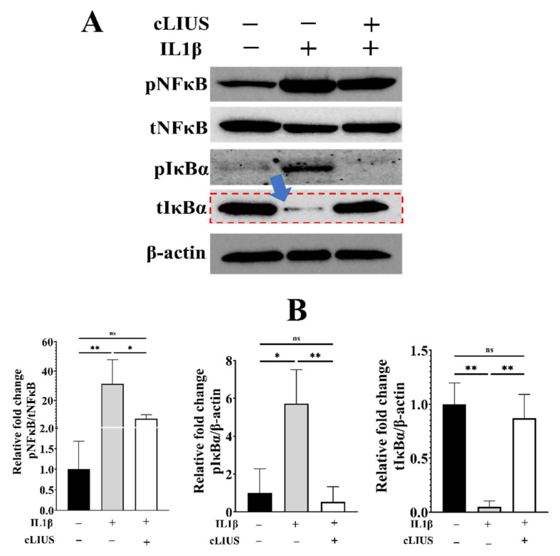
Proinflammatory joint environment, coupled with impeded chondrogenic differentiation of mesenchymal stromal cells (MSCs), led to inferior cartilage repair outcomes. Nuclear translocation of phosphorylated-NFκB downregulates SOX9 and hinders the chondrogenesis of MSCs. Strategies that minimize the deleterious effects of NFκB, while promoting MSC chondrogenesis, are of interest. This study establishes the ability of continuous low-intensity ultrasound (cLIUS) to preserve MSC chondrogenesis in a proinflammatory environment. MSCs were seeded in alginate:collagen hydrogels and cultured for 21 days in an ultrasound-assisted bioreactor (5.0 MHz, 2.5 Vpp; 4 applications/day) in the presence of IL1β and evaluated by qRT-PCR and immunofluorescence. The differential expression of markers associated with the NFκB pathway was assessed upon a single exposure of cLIUS and assayed by Western blotting, qRT-PCR, and immunofluorescence. Mitochondrial potential was evaluated by tetramethylrhodamine methyl ester (TMRM) assay. The chondroinductive potential of cLIUS was noted by the increased expression of SOX9 and COLII. cLIUS extended its chondroprotective effects by stabilizing the NFκB complex in the cytoplasm via engaging the IκBα feedback mechanism, thus preventing its nuclear translocation. cLIUS acted as a mitochondrial protective agent by restoring the mitochondrial potential and the mitochondrial mRNA expression in a proinflammatory environment. Altogether, our results demonstrated the potential of cLIUS for cartilage repair and regeneration under proinflammatory conditions.

Keywords: NFκB pathway; mesenchymal stromal cells; mitochondrial potential; ultrasound.


Conflict of interest statement

The authors declare no conflict of interest.

Figures

Figure 1
Figure 1
Experimental schematic. MSCs were seeded in alginate: collagen hydrogels or TCPs or coverslips, and divided into groups, as indicated. Group 1: cLIUS (−), IL1β (−); Group 2: IL1β (+), cLIUS (−); and Group 3: IL1β (+) cLIUS (+). Appropriate sample groups were treated with cytokine at a concentration of 10 ng/mL. cLIUS stimulation was applied as follows:14 kPa (5.0 MHz, 2.5 Vpp), and 10 or 20 min/application. Non-cytokine treated and non-cLIUS-stimulated samples served as controls. Upon completion of the study, samples were retrieved and subjected to the indicated outcome analyses.
Figure 2
Figure 2
Localization of pNFκB and COLII in hydrogel scaffolds. At the end of 21 days of culture, hydrogels were subjected to both gene expression and IF analysis. Briefly, hydrogels were fixed using 4% PFA in HBSSCM and stained against COLII and pNFκB (green fluorescence), respectively, in separate experiments (A,C), and nuclei were counter stained with DAPI (blue fluorescence). Z stacks of the hydrogels were captured using the Zeiss LSM 700 confocal microscope with 63× magnification (z step size 5 µm), and fluorescence intensity (B,D) was quantified using ImageJTM software (n = 30). Data are shown as the mean ± standard deviation of samples and p-value represents statistical significance (* p < 0.05; ** p < 0.01; **** p < 0.0001 and ns-nonsignificant) and scale bar represents 5 µm. ‘+’ indicates presence and ‘–’ indicates absence.
Figure 3
Figure 3
Gene expression analysis of MSCs in hydrogels. Cell homogenates (n = 10) were prepared from hydrogels; total RNA was extracted, and gene expression of lineage markers and catabolic markers was evaluated by qRT-PCR, and GAPDH was used as a housekeeping gene. Data are shown as the mean ± standard deviation of samples and p-value represents statistical significance (** p < 0.01; *** p < 0.001 and **** p < 0.0001).
Figure 4
Figure 4
Gene expression analysis in MSCs exposed to IL1β. MSCs were seeded on TCPs and treated as depicted in Figure 1. Homogenates from two wells per group served as one replicate, and three such replicates were used for gene expression analysis (n = 3). Total RNA was extracted, and the gene expression of anabolic and catabolic markers was evaluated by qRT-PCR; GAPDH was used as a housekeeping gene. Bar graph represents mean ± 95% confidence interval; p values indicate statistically significant differences (* p < 0.05; ** p < 0.01; **** p < 0.0001 and ns-nonsignificant).
Figure 5
Figure 5
Localization of SOX9 and pNFκB in MSCs exposed to IL1β. MSCs were seeded on coverslips and treated as depicted in Figure 1. Coverslips were fixed and double stained for pNFκB (green fluorescence) and SOX9 (red fluorescence) antibodies, and nuclei were counter stained with DAPI (blue fluorescence). (A) Images were captured using 63× magnification and presented. (B) Fluorescence intensity was quantified using ImageJTM software (n = 30). Bar graph represents mean ±95% confidence interval; p values indicate statistically significant differences. (**** p < 0.0001) and the scale bar represents 5 µm.
Figure 6
Figure 6
Phosphorylation of NFκB and IκBα. MSCs were seeded on TCP plates and treated as depicted in Figure 1. Total cell lysates were obtained and analyzed by Western blotting using specific antibodies. (A) Protein expression of phospho-NFκB, total NFκB, phospho-IκBα and total IκBα in indicated samples by Western blotting; β-actin was used as a loading control. (B) Blots were quantified using ImageJTM software and presented. Data are shown as the mean ± standard deviation of samples and p-value represents statistical significance (* p < 0.05; ** p < 0.01; and ns-nonsignificant). ‘+’ indicates presence and ‘–’ indicates absence.
Figure 7
Figure 7
Assessment of mitochondrial potential and mRNA expression under cLIUS. MSCs were seeded on coverslips and treated as depicted in Figure 1. Coverslips were treated with 100 nM TMRM reagent for 30 min, and live images were captured using the Zeiss LSM 700 confocal microscope, and images are presented in (A). Fluorescence data was quantified (n = 50) using ImageJTM software and the fluorescence intensity graph presented in (B) along with mitochondrial gene expression (C). All data shown as the mean ± standard deviation of samples and p-value represent statistical significance (**** p < 0.0001), and scale bar represents 100 µm. ‘+’ indicates presence and ‘–’ indicates absence.
Figure 8
Figure 8
Schematic representation of the cLIUS-induced chondroprotective mechanisms. cLIUS promoted MSC chondrogenesis by inhibiting the cytokine-induced activation of the NFκB signaling pathway by engaging the tIκBα feedback mechanism while upregulating the expression of SOX9, the collagen II transcription factor. It is also posited that cLIUS acts to preserve the mitochondrial potential, which is impacted by cytokines.

Similar articles

Cited by

References

    1. Cucchiarini M., Orth P., Rey-Rico A., Venkatesan J.K., Madry H. Current perspectives in stem cell research for knee cartilage repair. Stem Cells Cloning Adv. Appl. 2014;7:1–17. doi: 10.2147/SCCAA.S42880. - DOI - PMC - PubMed
    1. Gao J., Yao J.Q., Caplan A. Stem cells for tissue engineering of articular cartilage. Proc. Inst. Mech. Eng. Part H J. Eng. Med. 2007;221:441–450. doi: 10.1243/09544119JEIM257. - DOI - PubMed
    1. Martín A.R., Patel J., Zlotnick H.M., Carey J.L., Mauck R.L. Emerging therapies for cartilage regeneration in currently excluded ‘red knee’ populations. NPJ Regen. Med. 2019;4:12. doi: 10.1038/s41536-019-0074-7. - DOI - PMC - PubMed
    1. Steinert A.F., Rackwitz L., Gilbert F., Nöth U., Tuan R.S. Concise Review: The Clinical Application of Mesenchymal Stem Cells for Musculoskeletal Regeneration: Current Status and Perspectives. Stem Cells Transl. Med. 2012;1:237–247. doi: 10.5966/sctm.2011-0036. - DOI - PMC - PubMed
    1. Filardo G., Vannini F., Marcacci M., Andriolo L., Ferruzzi A., Giannini S., Kon E. Matrix-assisted autologous chondrocyte transplantation for cartilage regeneration in osteoarthritic knees: Results and failures at midterm follow-up. Am. J. Sports Med. 2012;41:95–100. doi: 10.1177/0363546512463675. - DOI - PubMed

Publication types