. 2022 Mar 12;12(3):438.
doi: 10.3390/biom12030438.

NAMPT Inhibitor and P73 Activator Represses P53 R175H Mutated HNSCC Cell Proliferation in a Synergistic Manner

Affiliations

NAMPT Inhibitor and P73 Activator Represses P53 R175H Mutated HNSCC Cell Proliferation in a Synergistic Manner

Bi-He Cai et al. Biomolecules. .

Abstract

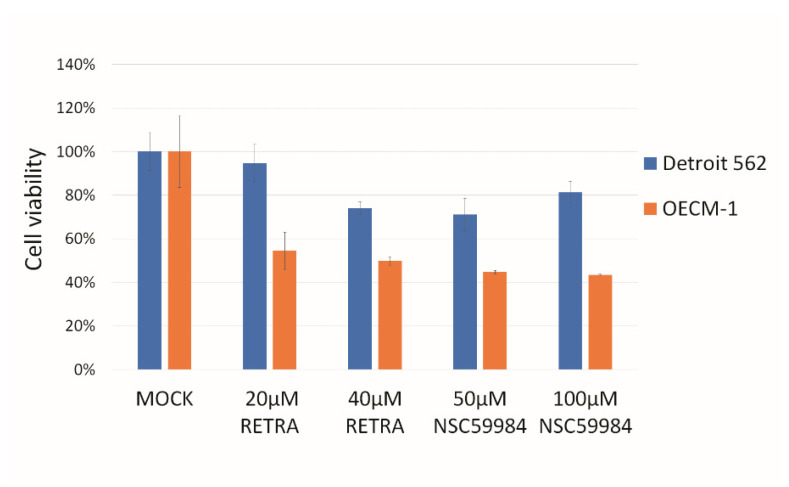
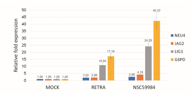
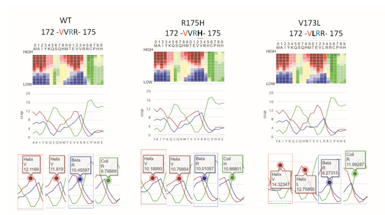
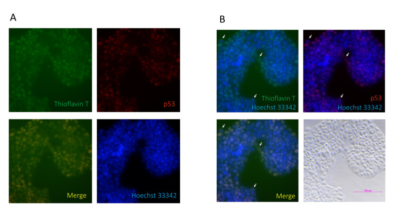
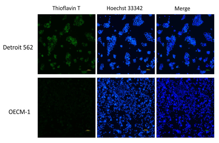
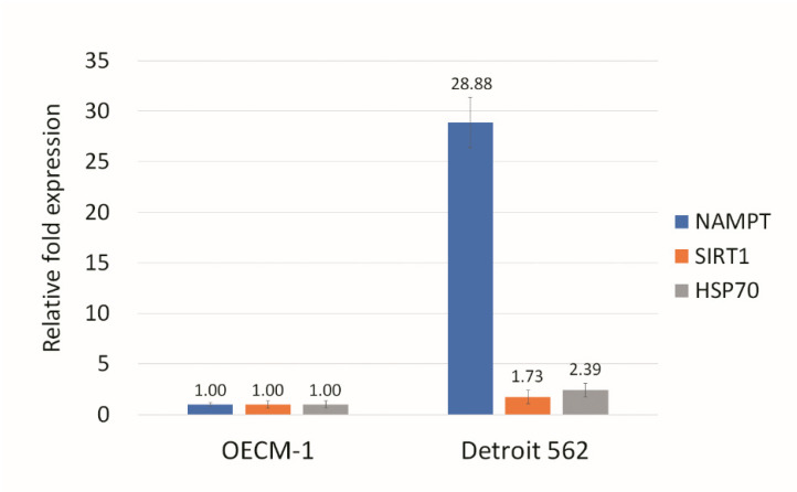
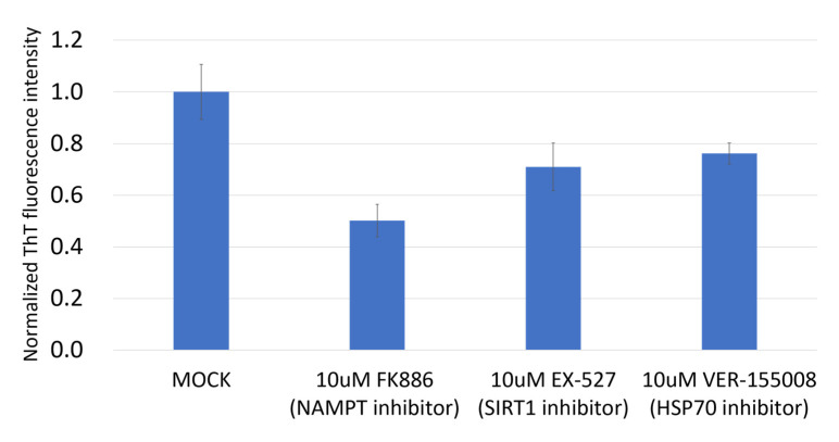
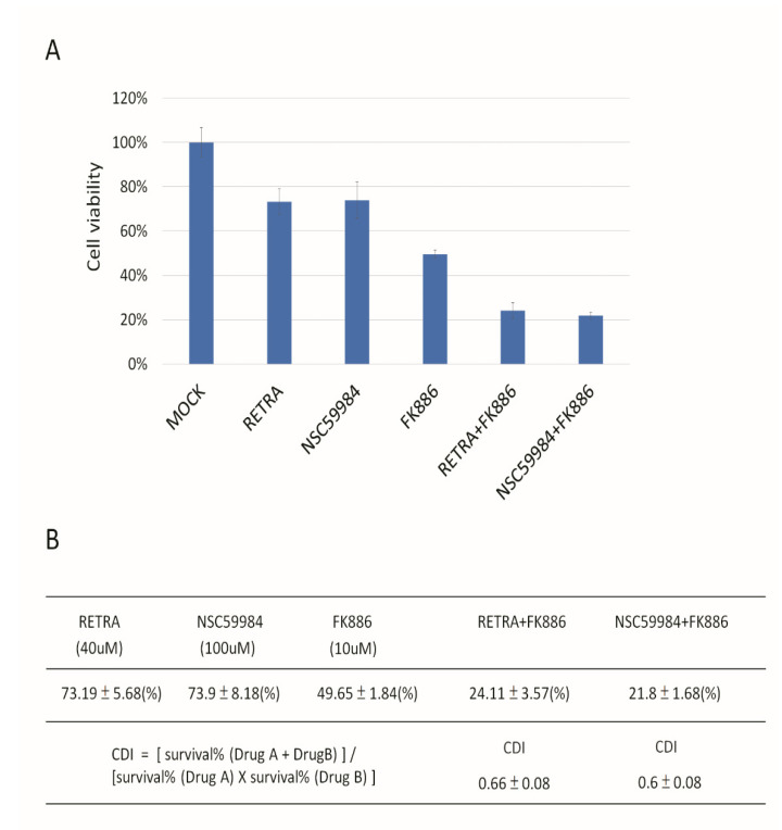
The p53 family has the following three members: p53, p63 and p73. p53 is a tumor suppressor gene that frequently exhibits mutation in head and neck cancer. Most p53 mutants are loss-of-function (LoF) mutants, but some acquire some oncogenic function, such as gain of function (GoF). It is known that the aggregation of mutant p53 can induce p53 GoF. The p73 activators RETRA and NSC59984 have an anti-cancer effect in p53 mutation cells, but we found that p73 activators were not effective in all head and neck squamous cell carcinoma (HNSCC) cell lines, with different p53 mutants. A comparison of the gene expression profiles of several regulator(s) in mutant HNSCC cells with or without aggregation of p53 revealed that nicotinamide phosphoribosyltransferase (NAMPT) is a key regulator of mutant p53 aggregation. An NAMPT inhibitor, to reduce abnormal aggregation of mutant p53, used in combination with a p73 activator, was able to effectively repress growth in HNSCC cells with p53 GoF mutants. This study, therefore, suggests a potential combination therapy approach for HNSCC with a p53 GoF mutation.

Keywords: NAMPT; aggregation; hand and neck; p53; p73.


Conflict of interest statement

The authors declare no conflict of interest.

Figures

Figure 1
Figure 1
p73 activators can repress HNSCC growth in HNSCC cell lines. Two p73 activators, RETRA and NSC59984, were able to repress OECM-1 cell growth and, to a lesser extent, Detroit 562 cell growth. Mock was calculated as 100% to normalize for other conditions.
Figure 2
Figure 2
p73 activator(s) activate p73 downstream genes in OCEM-1 cells. RETRA (40 μM) or NSC59984 (100 μM) could upregulate p73 downstream genes NEU4, JAG2, LIG1 and G6PD in OECM-1 cells. Mock (DMSO only) was calculated as 1 to normalize for other conditions.
Figure 3
Figure 3
The CSSP value of the β-strand of flanking amino acids (a.a.) on p53 V173L is lower than the wild-type (WT) and R175H. The propensity for each of the three secondary structure elements, α helix (red), β-strand (blue) and coil (green), is calculated at 20 different levels, so the sum of the CSSP scores is 20. R175H flanking a.a. reduces α-strand CSSP from 12.1169 (WT) to 10.18993. V173L flanking a.a. reduces β-strand CSSP from 10.45597 (WT) to 8.27313. V173L flanking a.a. has a 14.32347 α-strand CSSP larger than WT (12.1169) and R175H (10.18993).
Figure 4
Figure 4
p53 is colocalized with thioflavin T in Detroit 562 cells. (A) p53 was used to stain for protein aggregation and is shown in red; thioflavin T was used to stain for protein aggregation and is shown in green; Hoechst 33342 was used to stain for nuclear counterstains and is shown in blue. p53 signals are shown to have the same location as the thioflavin T signals in the merged figure. (B) The merged figure shows that there are some cells with cytosolic thioflavin T/p53 signals (arrows).
Figure 5
Figure 5
Protein aggregation in HNSCC cell lines. Thioflavin T was used to stain for protein aggregation and is shown in green; Hoechst 33342 was used to stain for nuclear counterstains and is shown in blue. Detroit 562 cells showed more thioflavin T signals than OECM1 cells.
Figure 6
Figure 6
NAMPT has a higher level of expression in Detroit 562 cells than in OECM-1 cells. Although the levels of NAMPT, SIRT1, and HSP70 expression in Detroit 562 were larger than in OECM-1, the NAMPT expression showed, by far, the greatest difference between the two cell lines. Gene expression in OECM1 cells was calculated as 1.
Figure 7
Figure 7
NAMPT inhibitor was able to repress protein aggregation signals. Although all the inhibitors tested (NAMPT, SIRT1 and HSP70) reduced the aggregation signals in Detroit 562, the NAMPT inhibitor had the most pronounced effect in reducing the protein aggregation. The protein aggregation signal was calculated as the OD ratio of ThT/Hoechst 33342 as normalized ThT fluorescence intensity. Mock was calculated as 1 to normalize for other conditions.
Figure 8
Figure 8
Synergistic repression of Detroit 562 cell growth mediated by p73 activators and NAMPT inhibitors. (A) RETRA, NSC59984 and NAMPT inhibitor (FK886) can inhibit Detroit 562 growth. Mock was calculated as 1 to normalize for other conditions. (B) The coefficient of drug interaction (CDI) was calculated as (A + B)/A×B. CDI < 1 = 1 or >1 indicates that the drugs are synergistic, additive or antagonistic, respectively. CDI < 0.7 indicates that the drug is significantly synergistic. p73 activator (RETRA) and NAMPT inhibitor (FK886) act synergistically (CDI = 0.66) to repress cell proliferation in Detroit 562 cells. Another p73 activator (NSC59984) and NAMPT inhibitor (FK886) also have synergic effects (CDI = 0.6) that reduce cell growth.

Similar articles

Cited by

References

    1. Levrero M., De Laurenzi V., Costanzo A., Gong J., Wang J.Y., Melino G. The p53/p63/p73 family of transcription factors: Overlapping and distinct functions. Pt 10J. Cell Sci. 2000;113:1661–1670. doi: 10.1242/jcs.113.10.1661. - DOI - PubMed
    1. Harms K.L., Chen X. The functional domains in p53 family proteins exhibit both common and distinct properties. Cell Death Differ. 2006;13:890–897. doi: 10.1038/sj.cdd.4401904. - DOI - PubMed
    1. Nigro J.M., Baker S.J., Preisinger A.C., Jessup J.M., Hostetter R., Cleary K., Bigner S.H., Davidson N., Baylin S., Devilee P. Mutations in the p53 gene occur in diverse human tumour types. Nature. 1989;342:705–708. doi: 10.1038/342705a0. - DOI - PubMed
    1. Shi C., Liu S., Tian X., Wang X., Gao P. A TP53 mutation model for the prediction of prognosis and therapeutic responses in head and neck squamous cell carcinoma. BMC Cancer. 2021;21:1035. doi: 10.1186/s12885-021-08765-w. - DOI - PMC - PubMed
    1. Pitolli C., Wang Y., Mancini M., Shi Y., Melino G., Amelio I. Do Mutations Turn p53 into an Oncogene? Int. J. Mol. Sci. 2019;20:6241. doi: 10.3390/ijms20246241. - DOI - PMC - PubMed

Publication types

MeSH terms

Substances