Review
. 2022 Mar 14;12(3):446.
doi: 10.3390/biom12030446.

Endogenous Human Proteins Interfering with Amyloid Formation

Affiliations
Review

Endogenous Human Proteins Interfering with Amyloid Formation

Anna L Gharibyan et al. Biomolecules. .

Abstract

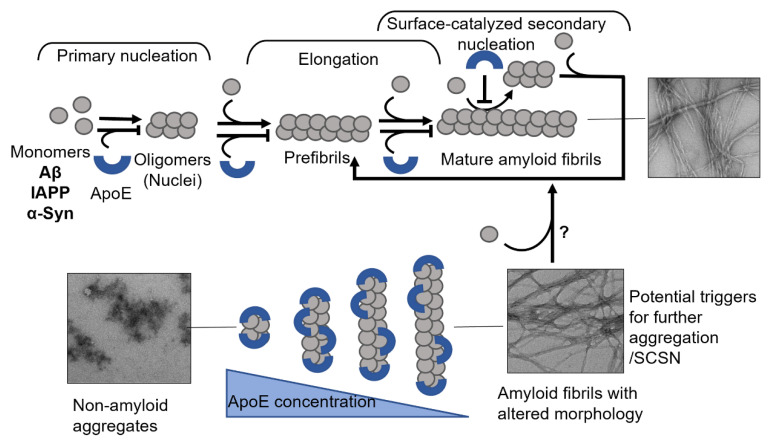
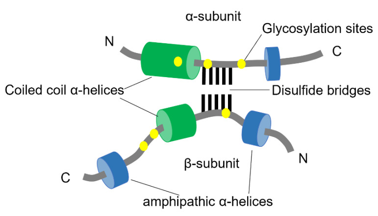
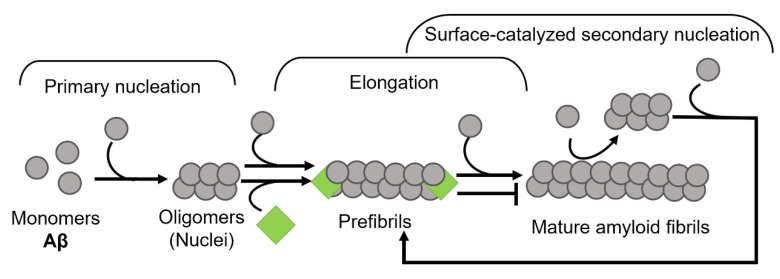
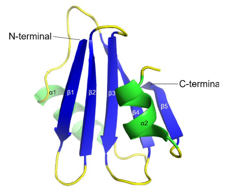
Amyloid formation is a pathological process associated with a wide range of degenerative disorders, including Alzheimer's disease, Parkinson's disease, and diabetes mellitus type 2. During disease progression, abnormal accumulation and deposition of proteinaceous material are accompanied by tissue degradation, inflammation, and dysfunction. Agents that can interfere with the process of amyloid formation or target already formed amyloid assemblies are consequently of therapeutic interest. In this context, a few endogenous proteins have been associated with an anti-amyloidogenic activity. Here, we review the properties of transthyretin, apolipoprotein E, clusterin, and BRICHOS protein domain which all effectively interfere with amyloid in vitro, as well as displaying a clinical impact in humans or animal models. Their involvement in the amyloid formation process is discussed, which may aid and inspire new strategies for therapeutic interventions.

Keywords: BRICHOS; IAPP; alpha-synuclein; amyloid inhibition; amyloid-beta; apolipoprotein E; clusterin; endogenous proteins; transthyretin.


Conflict of interest statement

The authors declare no conflict of interest.

Figures

Figure 1
Figure 1
Schematic models of amyloid fibril formation. (A) Linear polymerization, where partially unfolded or misfolded monomers assemble into short prefibrillar structures and by sequential addition of monomers elongate into mature fibrils. (B) Nucleation-dependent amyloid formation, where the monomers form soluble oligomeric nucleus during the lag phase, then assemble into larger prefibrillar structures followed by elongation and subsequently a saturation phase where mature fibrils are formed. Regarding some amyloid proteins, mature fibrils can serve as a template for the formation of new nuclei, resulting in a rate-enhancing process known as surface catalyzed secondary nucleation.
Figure 2
Figure 2
3D ribbon diagram of the native human TTR protein. (A). TTR monomer with eight β-strands A-H, and an α-helix between E and F strands. (B). TTR tetramer in two different orientations and indicated thyroxine-binding pocked. Key features are colored as β-strands in blue and α-helices in green. The figures are created from PDB-ID: 1DVQ.
Figure 3
Figure 3
Schematic presentation of suggested mechanisms for TTR interference with (A) Aβ, where TTR targets both primary and secondary nucleation and re-directs the reaction towards the formation of non-amyloid aggregates (modified from Nilsson et al., 2018 [37]), and (B) IAPP, where TTR specifically targets the elongation process (modified from Wasana Jayaweera et al., 2021 [91]) amyloid formation.
Figure 4
Figure 4
Full-length structure of Apolipoprotein E3 (PDB-ID: 2L7B). The N-terminal domain is colored in blue where the light-blue region is the LDL-receptor binding site, and the C-terminal domain is colored in green, where the light green region is the lipid-binding site. The red-colored residues at 112 and 158 positions are the varying residues between the three ApoE variants.
Figure 5
Figure 5
Mechanisms of ApoE interference with amyloid fibril formation. The schematic presentation in the upper panel shows that ApoE can target multiple steps of amyloid formation, including primary nucleation, elongation, and secondary nucleation. The lower panel shows the effect of ApoE concentration in interfering with amyloid formation process, where the higher concentration can convert the amyloid protein into non-amyloid aggregates and the lower concentrations lead to the production of amyloid fibrils with altered morphology.
Figure 6
Figure 6
Schematic structure of Clusterin. The two subunits of clusterin are assembled anti-parallel resulting in a heterodimeric molecule. The cysteine-rich centers are linked by five disulfide bridges (black lines) and are surrounded by two predicted coiled-coil α-helices (green) and three predicted amphipathic α-helices (blue). The N-linked glycosylation sites are indicated as yellow spots (adapted from Jones et al., 2002 [166]).
Figure 7
Figure 7
Schematic illustration of clusterin on the aggregation kinetics of Aβ. Clusterin perturbs the amyloid formation process by binding to fibril ends, thus inhibiting the elongation phase (modified from Scheidt et al., 2019 [186]).
Figure 8
Figure 8
3D ribbon backbone conformation of proSP-C BRICHOS domain (PDB-ID: 2YAD). The five β-strands (β1–β5) are shown in blue and the two α-helices (α1 and α2) in green.
Figure 9
Figure 9
Mechanism of BRICHOS domain in amyloid fibril formation. BRICHOS domain binds both to the ends as well as laterally on fibrils and thereby inhibits both elongation and secondary nucleation events (modified from Arosio et al., 2016 [215]).

Similar articles

Cited by

References

    1. Chiti F., Dobson C.M. Protein Misfolding, Amyloid Formation, and Human Disease: A Summary of Progress Over the Last Decade. Annu. Rev. Biochem. 2017;86:27–68. doi: 10.1146/annurev-biochem-061516-045115. - DOI - PubMed
    1. Bély M., Makovitzky J. Sensitivity and specificity of Congo red staining according to Romhányi. Comparison with Puchtler’s or Bennhold’s methods. Acta Histochem. 2006;108:175–180. doi: 10.1016/j.acthis.2006.03.017. - DOI - PubMed
    1. Frieden C. Protein aggregation processes: In search of the mechanism. Protein Sci. 2007;16:2334–2344. doi: 10.1110/ps.073164107. - DOI - PMC - PubMed
    1. Powers E., Powers D.L. The Kinetics of Nucleated Polymerizations at High Concentrations: Amyloid Fibril Formation Near and Above the “Supercritical Concentration”. Biophys. J. 2006;91:122–132. doi: 10.1529/biophysj.105.073767. - DOI - PMC - PubMed
    1. Hurshman A.R., White J.T., Powers E.T., Kelly J.W. Transthyretin Aggregation under Partially Denaturing Conditions Is a Downhill Polymerization. Biochemistry. 2004;43:7365–7381. doi: 10.1021/bi049621l. - DOI - PubMed

Publication types