. 2022 Mar 15;12(3):455.
doi: 10.3390/biom12030455.

Inherited Retinal Degeneration: PARP-Dependent Activation of Calpain Requires CNG Channel Activity

Affiliations

Inherited Retinal Degeneration: PARP-Dependent Activation of Calpain Requires CNG Channel Activity

Jie Yan et al. Biomolecules. .

Abstract

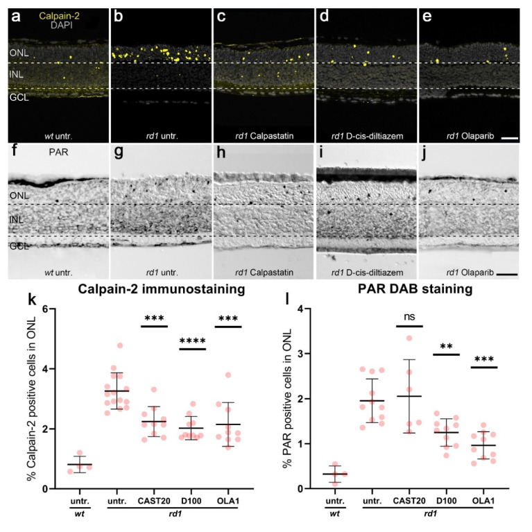
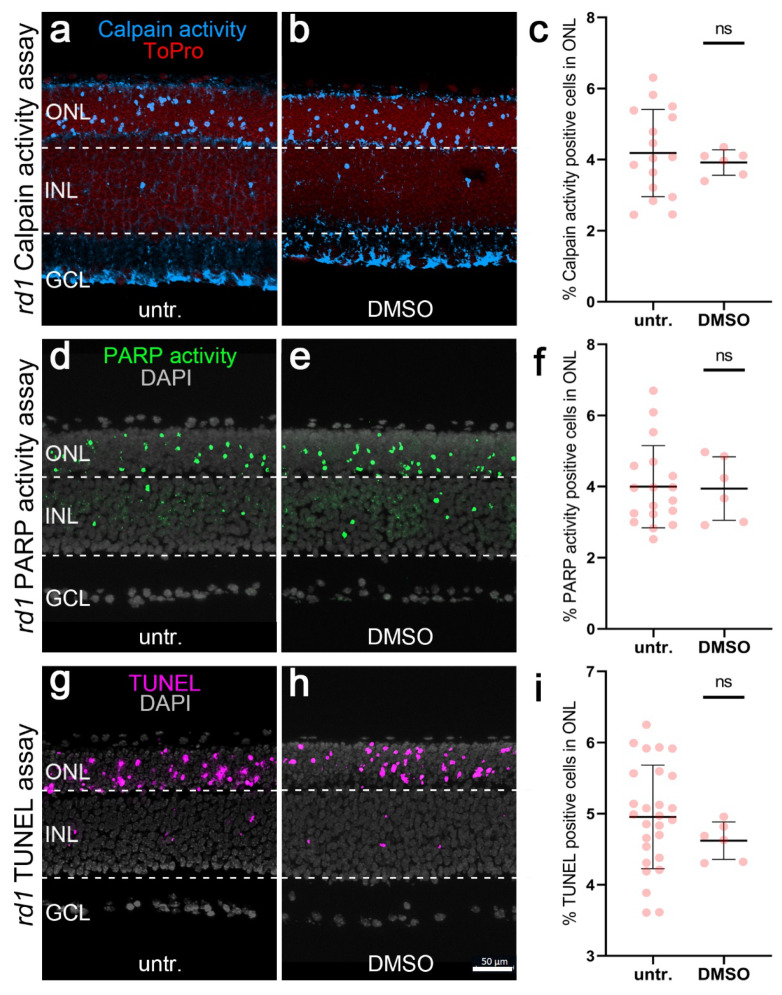
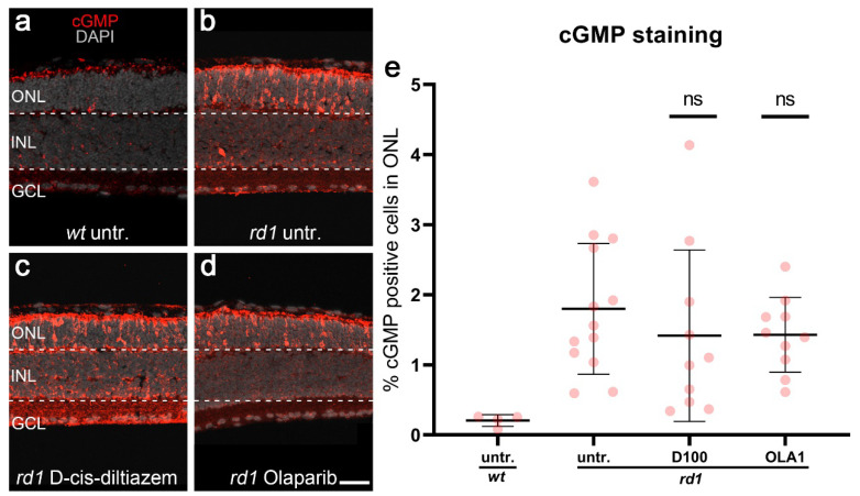
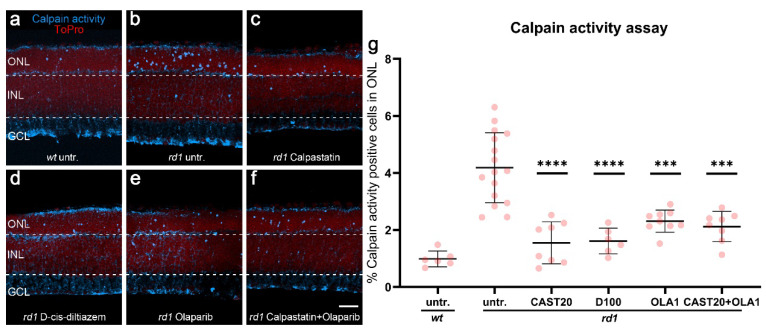
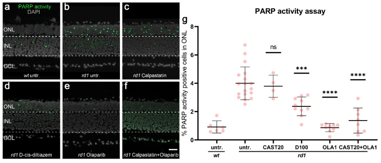
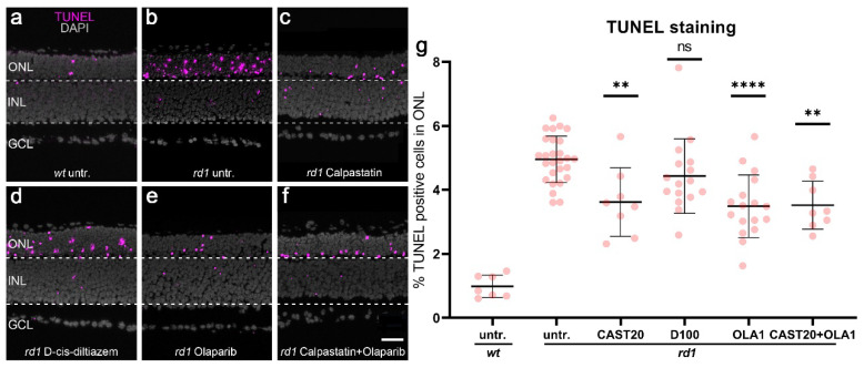
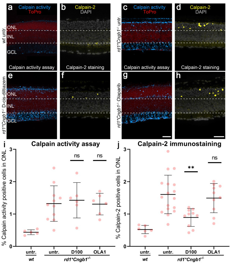
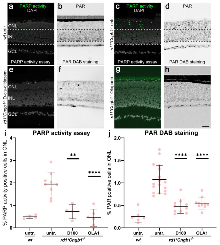
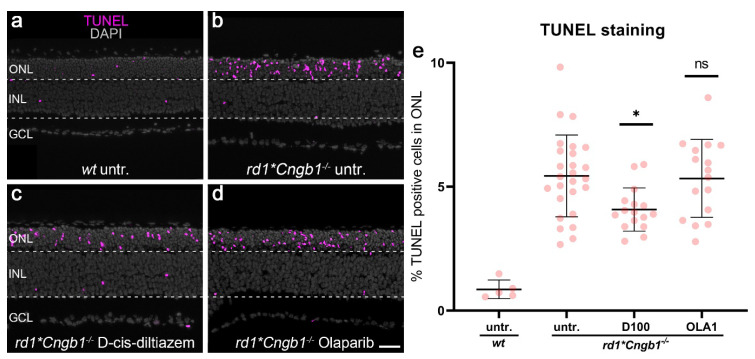
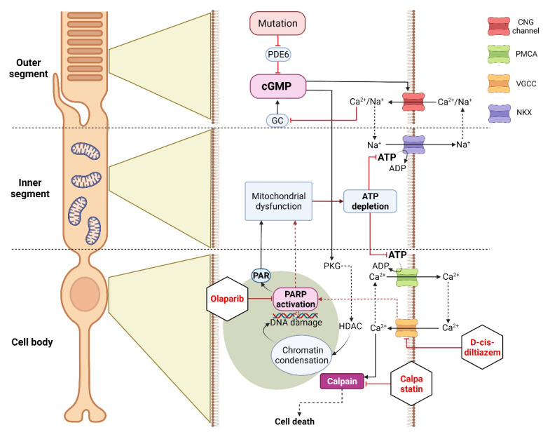
Inherited retinal degenerations (IRDs) are a group of blinding diseases, typically involving a progressive loss of photoreceptors. The IRD pathology is often based on an accumulation of cGMP in photoreceptors and associated with the excessive activation of calpain and poly (ADP-ribose) polymerase (PARP). Inhibitors of calpain or PARP have shown promise in preventing photoreceptor cell death, yet the relationship between these enzymes remains unclear. To explore this further, organotypic retinal explant cultures derived from wild-type and IRD-mutant mice were treated with inhibitors specific for calpain, PARP, and voltage-gated Ca2+ channels (VGCCs). The outcomes were assessed using in situ activity assays for calpain and PARP and immunostaining for activated calpain-2, poly (ADP-ribose), and cGMP, as well as the TUNEL assay for cell death detection. The IRD models included the Pde6b-mutant rd1 mouse and rd1*Cngb1-/- double-mutant mice, which lack the beta subunit of the rod cyclic nucleotide-gated (CNG) channel and are partially protected from rd1 degeneration. We confirmed that an inhibition of either calpain or PARP reduces photoreceptor cell death in rd1 retina. However, while the activity of calpain was decreased by the inhibition of PARP, calpain inhibition did not alter the PARP activity. A combination treatment with calpain and PARP inhibitors did not synergistically reduce cell death. In the slow degeneration of rd1*Cngb1-/- double mutant, VGCC inhibition delayed photoreceptor cell death, while PARP inhibition did not. Our results indicate that PARP acts upstream of calpain and that both are part of the same degenerative pathway in Pde6b-dependent photoreceptor degeneration. While PARP activation may be associated with CNG channel activity, calpain activation is linked to VGCC opening. Overall, our data highlights PARP as a target for therapeutic interventions in IRD-type diseases.

Keywords: HDAC; PKG; cGMP; calcium; nonapoptotic cell death; photoreceptor degeneration; retinitis pigmentosa.


Conflict of interest statement

The authors declare no conflict of interest.

Figures

Scheme A1
Scheme A1
Effects of calpastatin, D-cis-diltiazem, and Olaparib on calpain-2 activation and PAR. Calpain-2 immunostaining (yellow; ae) and PAR DAB staining (black, fj) were performed on wt and rd1 retina. DAPI (grey) was used as nuclear counterstaining. Untreated rd1 retina (untr.; b,g) was compared to retina treated with calpastatin (c,h), D-cis-diltiazem (d,i), and Olaparib (e,j), respectively. The scatter plots show the percentages of ONL-positive cells for calpain-2 (k) and PAR (l) in wt and treated rd1 retina, compared with rd1 control (untr.). Statistical significance was assessed using one-way ANOVA, and Tukey’s multiple comparison post hoc testing was performed between the control and 20-μM calpastatin (CAST20), 100-μM D-cis-diltiazem (D100), and 1-μM Olaparib (OLA1). In rd1 retina, all treatments decreased the numbers of cells positive for calpain-2, while the number of PAR-positive cells were not reduced by CAST20. In calpain-2 immunostaining: Untr. wt: 4 explants from 2 different mice; untr. rd1: 15/15; CAST20 rd1: 10/10; D100 rd1: 10/10; OLA1 rd1: 10/10. In PAR DAB staining: untr. wt: 4/2; untr. rd1: 16/16; CAST20 rd1: 6/6; D100 rd1: 10/10; OLA1 rd1: 10/10; error bars represent SD; ns = p > 0.05, ** = p ≤ 0.01, *** = p ≤ 0.001, and **** = p ≤ 0.0001. ONL = outer nuclear layer, INL = inner nuclear layer, GCL = ganglion cell layer. Scale bar = 50 µm.
Scheme A2
Scheme A2
Effect of DMSO on calpain activity, PARP activity, and TUNEL staining in rd1 retinal explants. Calpain activity (blue), PARP activity (green), and TUNEL (magenta) were used in rd1 retinal explant cultures. ToPro (red) and DAPI (grey) were used as nuclear counterstains. rd1 control retina (untr.; a,d,g) was compared to retina treated with 0.1% DMSO (DMSO; b,e,h). There was no difference between positive cells detected in the rd1 outer nuclear layer (ONL), with or without DMSO. The scatter plot (e) shows the percentage of calpain activity (c), PARP activity (f), and TUNEL-positive cells (i). In the calpain activity assay, untr. rd1: 16 explants derived from 16 different animals; DMSO rd1: 6/6. In the PARP activity assay, untr. rd1: 18/18; DMSO rd1: 6/6. In the TUNEL assay, untr. rd1: 27/27; DMSO rd1: 6/6; error bars represent SD; ns = p > 0.05, INL = inner nuclear layer, GCL = ganglion cell layer. Scale bar = 50 µm.
Scheme A3
Scheme A3
Photoreceptor accumulation of cGMP in different experimental conditions. cGMP immunostaining (red) was used in wt and rd1 retinal explant cultures. DAPI (grey) was used as a nuclear counterstain. In wt retina (untr.; a), only a few cells in the outer nuclear layer (ONL) were labeled cGMP-positive. rd1 control retina (untr.; b) was compared to retina treated with either 100-µM D-cis-diltiazem (D100, c) or 1-µM Olaparib (OLA1, d). Statistical significance was assessed using one-way ANOVA, and Tukey’s multiple comparison post hoc testing was performed between control, 100-μM D-cis-diltiazem (D100), and 1-μM Olaparib (OLA1). Note the large numbers of cGMP expressed in the rd1 ONL. The scatter plot (e) shows the percentage of cGMP-positive cells in the ONL. Neither D-cis-diltiazem or Olaparib reduced the cGMP-positive cells. Untr. wt: 4 explants from 2 different mice; untr. rd1: 13/13; D100 rd1: 10/10; OLA1 rd1: 10/10; error bars represent SD; ns = p > 0.05. INL = inner nuclear layer, GCL = ganglion cell layer. Scale bar = 50 µm.
Figure 1
Figure 1
Effects of calpastatin, D-cis-diltiazem, Olaparib, and combination treatments on calpain activity. The calpain activity assay (blue) was performed on unfixed wt (a) and rd1 retinal cross-sections. ToPro (red) was used as nuclear counterstaining. Untreated rd1 retina (untr.; b) was compared to retina treated with either calpastatin (c), D-cis-diltiazem (d), Olaparib (e), or calpastatin and Olaparib combined (f). The scatter plots show the percentages of outer nuclear layer (ONL) cells positive for calpain activity (g) in wt and rd1 retina. Statistical significance was assessed using one-way ANOVA and Tukey’s multiple comparison post hoc testing performed between the control (rd1 untreated) and 20-μM calpastatin (CAST20), 100-μM D-cis-diltiazem (D100), 1-μM Olaparib (OLA1), and 20-μM calpastatin combined with 1-μM Olaparib (CAST20+OLA1). All treatments reduced the calpain activity in rd1 ONL; however, there was no added synergistic benefit from the CAST20+OLA1 combination. Untr. wt: 6 explants from 3 different mice; untr. rd1: 16/16; CAST20 rd1: 8/8; D100 rd1: 6/6; OLA1 rd1: 9/9; CAST20+OLA1 rd1: 8/8; error bars represent SD; *** = p ≤ 0.001 and **** = p ≤ 0.0001. INL = inner nuclear layer, GCL = ganglion cell layer. Scale bar = 50 µm.
Figure 2
Figure 2
Effects of calpastatin, D-cis-diltiazem, Olaparib, and combination treatments on PARP activity. PARP activity assay (green) in wt and rd1 retina. DAPI (grey) was used as nuclear counterstaining. Untreated wt (untr.; a) was compared to untreated rd1 retina (b) and retinae treated with either calpastatin (c), D-cis-diltiazem (d), Olaparib (e), or calpastatin and Olaparib combined (f). The scatter plots show the percentages of the outer nuclear layer (ONL) cells positive for PARP activity (g) in wt and rd1 retina. Statistical significance was assessed using one-way ANOVA and Tukey’s multiple comparison post hoc testing performed between the control (rd1 untreated) and 20-μM calpastatin (CAST20), 100-μM D-cis-diltiazem (D100), 1-μM Olaparib (OLA1), and 20-μM calpastatin combined with 1-μM Olaparib (CAST20+OLA1). Calpastatin did not reduce the PARP activity, while D-cis-diltiazem and Olaparib did. Untr. wt: 6 explants from 3 different mice; untr. rd1: 18/18; CAST20 rd1: 4/4; D100 rd1: 10/10; OLA1 rd1: 9/9; CAST20+OLA1 rd1: 8/8; error bars represent SD; ns = p > 0.05, *** = p ≤ 0.001, and **** = p ≤ 0.0001. INL = inner nuclear layer, GCL = ganglion cell layer. Scale bar = 50 µm.
Figure 3
Figure 3
Effects of calpastatin, D-cis-diltiazem, Olaparib, and combination treatments on rd1 retinal cell viability. The TUNEL assay labeled dying cells (magenta) in wt and rd1 retinal explant cultures. DAPI (grey) was used as a nuclear counterstain. Untreated wt (a) and rd1 control retina (untr.; b) were compared to retina treated with either 20-µM calpastatin (CAST20, c), 100-µM D-cis-diltiazem (D100, d), 1-µM Olaparib (OLA1, e), or 20-µM calpastatin combined with 1-µM Olaparib (CAST20+OLA1, f). Note the large numbers of dying cells in the rd1 outer nuclear layer (ONL). The scatter plot (g) shows the percentage of TUNEL-positive cells in the ONL. Statistical significance was assessed using one-way ANOVA and Tukey’s multiple comparison post hoc testing performed between the control (rd1 untreated) and 20-μM calpastatin (CAST20), 100-μM D-cis-diltiazem (D100), 1-μM Olaparib (OLA1), and 20-μM calpastatin combined with 1-μM Olaparib (CAST20+OLA1). Except for D-cis-diltiazem, all treatments decreased rd1 retinal degeneration. The combination treatment CAST20+OLA1 did not improve the therapeutic effect seen with the respective single treatments. Untr. wt: 7 explants from 4 different mice; untr. rd1: 27/27; CAST20 rd1: 8/8; D100 rd1: 16/16; OLA1 rd1: 17/17; CAST20+OLA1 rd1: 8/8; error bars represent SD; ns = p > 0.05, ** = p ≤ 0.01, and **** = p ≤ 0.0001. ONL = outer nuclear layer, INL = inner nuclear layer, GCL = ganglion cell layer. Scale bar = 50 µm.
Figure 4
Figure 4
Effects of D-cis-diltiazem and Olaparib on calpain activity in rd1*Cngb1−/− retina. The calpain activity assay (blue) and an immunostaining for activated calpain-2 (yellow) were performed on wt (a,b) and rd1*Cngb1−/− retina. DAPI (grey) was used as nuclear counterstaining. Untreated rd1*Cngb1−/− retina (untr.; c,d) was compared to retina treated with D-cis-diltiazem (e,f) or Olaparib (g,h). The scatter plots show the percentages of ONL-positive cells for calpain activity (i) and activated calpain-2 (j) in the wt and treated rd1*Cngb1−/− retina compared with the rd1*Cngb1−/− control (untr.). Statistical significance was assessed using one-way ANOVA and Tukey’s multiple comparison post hoc testing performed between the control (rd1*Cngb1−/− untreated), 100-μM D-cis-diltiazem (D100), and 1-μM Olaparib (OLA1). In rd1*Cngb1−/−, only D-cis-diltiazem reduced the cells positive for activated calpain-2. In the calpain activity assay, untr. wt: 5 explants from 3 different mice; untr. rd1*Cngb1−/−: 11/11; D100 rd1*Cngb1−/−: 6/6; OLA1 rd1*Cngb1−/−: 6/6; in calpain-2 immunostaining, untr. wt: 6/3; untr. rd1*Cngb1−/−: 17/17; D100 rd1*Cngb1−/−: 10/10; OLA1 rd1*Cngb1−/−: 10/10; error bars represent SD; ns = p > 0.05 and ** = p ≤ 0.01. ToPro (red) and ONL = outer nuclear layer, INL = inner nuclear layer, GCL = ganglion cell layer. Scale bar = 50 µm.
Figure 5
Figure 5
Effects of D-cis-diltiazem and Olaparib on PARP activity and PAR accumulation in rd1*Cngb1−/− double-mutant retina. The PARP activity assay (green) and immunostaining for PAR (black) were performed on wt (a,b) and rd1*Cngb1−/− retina (ch). DAPI (grey) was used as nuclear counterstaining. Untreated rd1*Cngb1−/− retina (untr.; c,d) was compared to retina treated with D-cis-diltiazem (e,f) or Olaparib (g,h). The scatter plots show the percentages of outer nuclear layer (ONL) cells positive for PARP activity (i) and PAR (j) in wt and treated rd1*Cngb1−/− retina compared to the rd1*Cngb1−/− control (untr.). Statistical significance was assessed using one-way ANOVA and Tukey’s multiple comparison post hoc testing performed between the control (rd1*Cngb1−/− untreated) and 100-μM D-cis-diltiazem (D100) or 1-μM Olaparib (OLA1). D-cis-diltiazem strongly decreased the PARP activity and PAR. In the PARP activity assay, untr. wt: 4 explants from 2 different mice; untr. rd1*Cngb1−/−: 9/9; D100 rd1*Cngb1−/−: 4/4; OLA1 rd1*Cngb1−/−: 6/6. In PAR DAB staining, untr. wt: 6/3; untr. rd1*Cngb1−/−: 17/17; D100 rd1*Cngb1−/−: 10/10; OLA1 rd1*Cngb1−/−: 10/10; error bars represent SD; ** = p ≤ 0.01 and **** = p ≤ 0.0001. INL = inner nuclear layer, GCL = ganglion cell layer. Scale bar = 50 µm.
Figure 6
Figure 6
Effects of D-cis-diltiazem and Olaparib on rd1*Cngb1−/− retinal cell viability. The TUNEL assay labeled dying cells (magenta) in wild-type (wt) and rd1*Cngb1−/− retinal explant cultures. DAPI (grey) was used as a nuclear counterstain. (a) In wt retina, only a small fraction of cells in the outer nuclear layer (ONL) were TUNEL-positive. (b) Untreated (untr.) rd1*Cngb1−/− double-mutant retina was compared to retina treated with either 100-µM D-cis-diltiazem (D100, (c) or 1-µM Olaparib (OLA1, (d,e) The scatter plot shows the percentage of TUNEL-positive cells. Statistical significance was assessed using one-way ANOVA and Tukey’s multiple comparison post hoc testing performed between the control (rd1*Cngb1−/− untreated) and 20-μM calpastatin (CAST20), 100-μM D-cis-diltiazem (D100), 1-μM Olaparib (OLA1), and 20-μM calpastatin combined with 1-μM Olaparib (CAST20+OLA1). Only D-cis-diltiazem alleviated the rd1*Cngb1−/− retinal degeneration. Untr. wt: 5 explants from 3 different mice; untr. rd1*Cngb1−/−: 26/26; D100 rd1*Cngb1−/−: 16/16; OLA1 rd1*Cngb1−/−: 16/16; error bars represent SD; ns = p > 0.05 and * = p ≤ 0.05. INL = inner nuclear layer, GCL = ganglion cell layer. Scale bar = 50 µm.
Figure 7
Figure 7
Differential effects of experimental conditions on cGMP-dependent cell death in rd1 photoreceptors. The mutation-induced cGMP accumulation activates cyclic nucleotide-gated (CNG) channels in the outer segment, leading to Na+-and Ca2+-influx and photoreceptor depolarization. This leads to opening of voltage-gated Ca2+-channels (VGCCs) in the cell body, causing further Ca2+- influx. In the cell body, high Ca2+ levels may activate calpain if not controlled by ATP-dependent plasma membrane Ca2+-ATPase (PMCA). In addition, cGMP-dependent activation of protein kinase G (PKG) has been associated with histone-deacetylase (HDAC) activity, causing chromatin condensation and DNA breaks, which may trigger PARP activation. Excessive consumption of NAD+ by PARP and the production of PAR may cause mitochondrial dysfunction, leading to ATP shortage. Calpastatin treatment blocks calpain activation, decreasing proteolytic damage to the cell, even in the presence of CNG channel/VGCC-mediated Ca2+-influx. D-cis-diltiazem inhibits VGCCs in the cell body, reducing intracellular Ca2+-levels and calpain activity. Moreover, VGCCs could be involved in PARP activation, even though D-cis-diltiazem fails to delay rd1 rod degeneration. Olaparib blocks PARP activity, decreasing NAD+ consumption and PAR generation. This may preserve mitochondrial function and intracellular ATP levels, allowing PMCA to extrude Ca2+ and keeping calpain activity low.

Similar articles

Cited by

References

    1. Duncan J.L., Pierce E.A., Laster A.M., Daiger S.P., Birch D.G., Ash J.D., Iannaccone A., Flannery J.G., Sahel J.A., Zack D.J., et al. Technology, Inherited retinal degenerations: Current landscape and knowledge gaps. Transl. Vis. Sci. Technol. 2018;74:6. doi: 10.1167/tvst.7.4.6. - DOI - PMC - PubMed
    1. Bertelsen M., Jensen H., Bregnhøj J.F., Rosenberg T. Prevalence of generalized retinal dystrophy in Denmark. Ophthalmic Epidemiol. 2014;21:217–223. doi: 10.3109/09286586.2014.929710. - DOI - PubMed
    1. Hartong D.T., Berson E.L., Dryja T.P. Retinitis pigmentosa. Lancet. 2006;368:1795–1809. doi: 10.1016/S0140-6736(06)69740-7. - DOI - PubMed
    1. Sahel J.A., Marazova K., Audo I. Clinical characteristics and current therapies for inherited retinal degenerations. Cold Spring Harb. Perspect. Med. 2015;5:a017111. doi: 10.1101/cshperspect.a017111. - DOI - PMC - PubMed
    1. Arango-Gonzalez B., Trifunović D., Sahaboglu A., Kranz K., Michalakis S., Farinelli P., Koch S., Koch F., Cottet S., Janssen-Bienhold U., et al. Identification of a common non-apoptotic cell death mechanism in hereditary retinal degeneration. PLoS ONE. 2014;9:e112142. doi: 10.1371/journal.pone.0112142. - DOI - PMC - PubMed

Publication types

MeSH terms

LinkOut - more resources