Review
. 2022 Mar 18;12(3):467.
doi: 10.3390/biom12030467.

Acetylation, Phosphorylation, Ubiquitination (Oh My!): Following Post-Translational Modifications on the Ubiquitin Road

Affiliations
Review

Acetylation, Phosphorylation, Ubiquitination (Oh My!): Following Post-Translational Modifications on the Ubiquitin Road

Rachel E Lacoursiere et al. Biomolecules. .

Abstract

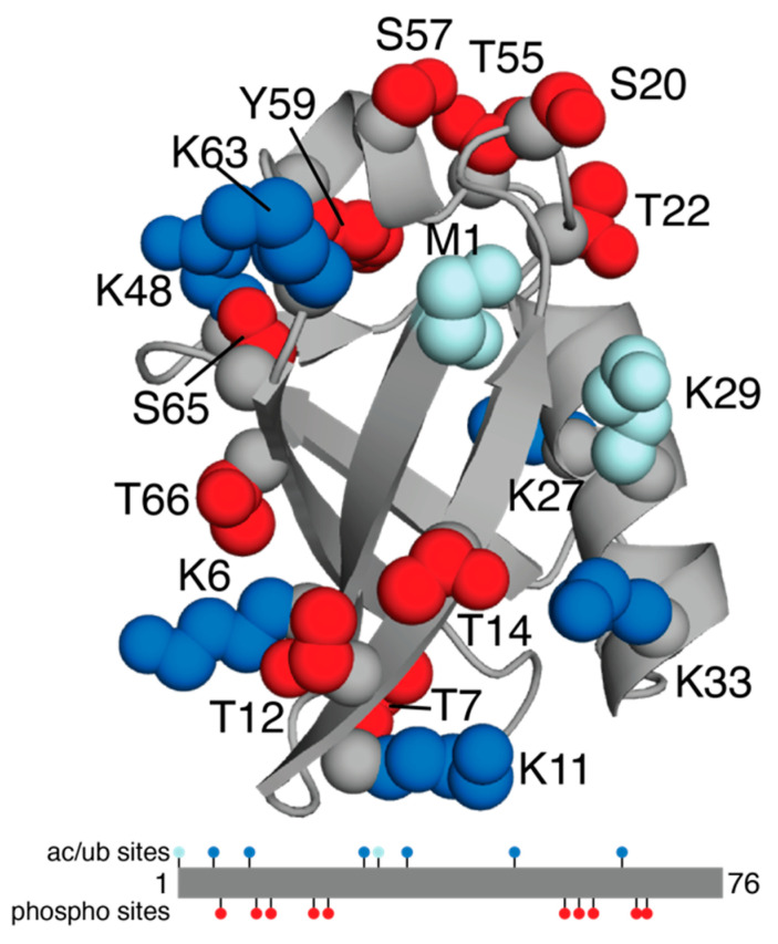
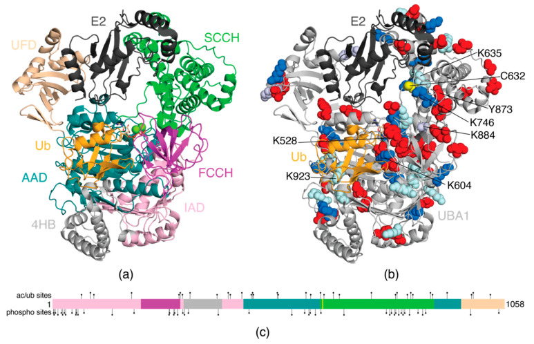
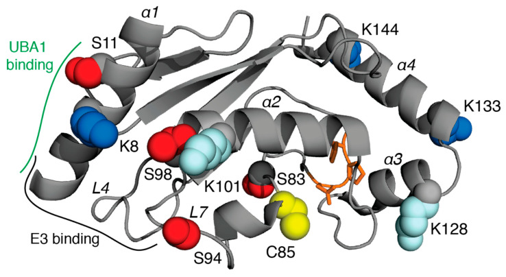
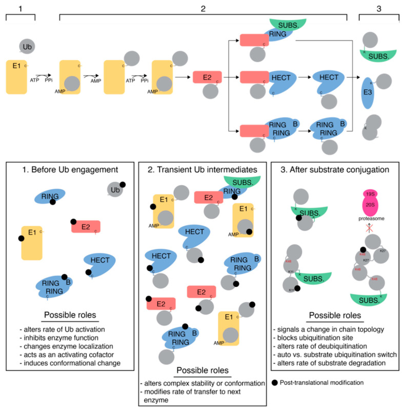
Ubiquitination is controlled by a series of E1, E2, and E3 enzymes that can ligate ubiquitin to cellular proteins and dictate the turnover of a substrate and the outcome of signalling events such as DNA damage repair and cell cycle. This process is complex due to the combinatorial power of ~35 E2 and ~1000 E3 enzymes involved and the multiple lysine residues on ubiquitin that can be used to assemble polyubiquitin chains. Recently, mass spectrometric methods have identified that most enzymes in the ubiquitination cascade can be further modified through acetylation or phosphorylation under particular cellular conditions and altered modifications have been noted in different cancers and neurodegenerative diseases. This review provides a cohesive summary of ubiquitination, acetylation, and phosphorylation sites in ubiquitin, the human E1 enzyme UBA1, all E2 enzymes, and some representative E3 enzymes. The potential impacts these post-translational modifications might have on each protein function are highlighted, as well as the observations from human disease.

Keywords: acetylation; cancer; neurodegenerative disease; phosphorylation; protein structure; proteomics; ubiquitination.


Conflict of interest statement

The authors declare no conflict of interest.

Figures

Figure 1
Figure 1
Ubiquitin post-translational modifications. The cartoon structure of Ub (PDB 1UBQ) [91] is shown in grey with PTM sites indicated: ubiquitination only (pale blue), acetylation or ubiquitination (blue), and phosphorylation (red). Also shown is the linear structure of Ub with the PTMs.
Figure 2
Figure 2
Cartoon model of the human E1 enzyme UBA1 in complex with the E2 conjugating enzyme, Ubc4 and ubiquitin. (a) Domains in UBA1 (PDB 6DC6) are indicated as 4HB (light grey), IAD (light pink), AAD (teal), FCCH (magenta), SCCH (green), UFD (wheat). The E2 enzyme (dark grey) and bound ubiquitin (orange) at the adenylation site are also shown. The E2 enzyme was modeled based on PDB coordinates 4II2 [98] and occupies a position consistent with other E1:E2 structures [99,100,101]. (b) The cartoon structure of UBA1 is shown in light grey, and the locations of acetylation or ubiquitination (blue), acetylation only (light purple), ubiquitination only (pale blue), and phosphorylation (red) sites in UBA1 are indicated. The catalytic cysteine (C632) is shown in yellow, and the E2 and Ub-adenylate are coloured as in (a). Only those PTM sites discussed in detail in the text are labelled here. (c) Linear domain organization of human UBA1 with colours matched to (a). The catalytic cysteine is indicated by a yellow bar, and all observed PTM sites in human UBA1 are indicated to the top (acetylation or ubiquitination) and bottom (phosphorylation) of the schematic.
Figure 3
Figure 3
Acetylation, phosphorylation, and ubiquitination sites in E2 conjugating enzymes. Residues observed to be acetylated or ubiquitinated (blue), phosphorylated (red), or only ubiquitinated (pale blue) in the UBE2D family (UBE2D1-UBE2D4) are indicated using the structure of UBE2D3 (PDB 5EGG) as a template [118]. Not all sites are found in all family members. The catalytic cysteine (C85) is shown in yellow and the HPN motif is shown in orange sticks. Helices and discussed loops are labelled in italics. Regions along helix α1 and the L4, L7 loops where UBA1 and E3 enzymes interact are indicated.
Figure 4
Figure 4
Implications of post-translational modification of ubiquitination proteins. The ubiquitination pathway for RING, HECT, and RBR E3 ligases is shown at the top. Post-translational modification of these proteins can occur at three points in the ubiquitination cycle: (1) before Ub engagement, (2) contained in a transient Ub complex, or (3) after substrate conjugation. Thioester intermediates are indicated by ‘C’ (cysteine), while stable isopeptide linkages are denoted by ‘K’ (lysine). Potential roles of PTMs at each point in the cascade are listed.

References

    1. Ye Y., Rape M. Building Ubiquitin Chains: E2 Enzymes at Work. Nat. Rev. Mol. Cell Biol. 2009;10:755–764. doi: 10.1038/nrm2780. - DOI - PMC - PubMed
    1. Ali I., Conrad R.J., Verdin E., Ott M. Lysine Acetylation Goes Global: From Epigenetics to Metabolism and Therapeutics. Chem. Rev. 2018;118:1216–1252. doi: 10.1021/acs.chemrev.7b00181. - DOI - PMC - PubMed
    1. Buuh Z.Y., Lyu Z., Wang R.E. Interrogating the Roles of Post-Translational Modifications of Non-Histone Proteins: Miniperspective. J. Med. Chem. 2018;61:3239–3252. doi: 10.1021/acs.jmedchem.6b01817. - DOI - PubMed
    1. Rittinger K., Ikeda F. Linear Ubiquitin Chains: Enzymes, Mechanisms and Biology. Open Biol. 2017;7:170026. doi: 10.1098/rsob.170026. - DOI - PMC - PubMed
    1. Hermida M.A., Dinesh Kumar J., Leslie N.R. GSK3 and Its Interactions with the PI3K/AKT/MTOR Signalling Network. Adv. Biol. Regul. 2017;65:5–15. doi: 10.1016/j.jbior.2017.06.003. - DOI - PubMed

Publication types