

Pleuropulmonary MDCT Findings: Comparison between Children with Pulmonary Vein Stenosis and Prematurity-Related Lung Disease
- PMID: 35327727
- PMCID: PMC8947577
- DOI: 10.3390/children9030355
Pleuropulmonary MDCT Findings: Comparison between Children with Pulmonary Vein Stenosis and Prematurity-Related Lung Disease
Abstract
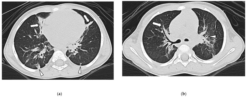
Purpose: To retrospectively compare the pleuropulmonary MDCT findings in children with pulmonary vein stenosis (PVS) and prematurity-related lung disease (PLD). Materials and Methods: All consecutive infants and young children (≤18 years old) who underwent thoracic MDCT studies from July 2004 to November 2021 were categorized into two groups—children with PVS (Group 1) and children with PLD without PVS (Group 2). Two pediatric radiologists independently evaluated thoracic MDCT studies for the presence of pleuropulmonary abnormalities as follows—(1) in the lung (ground-glass opacity (GGO), triangular/linear plaque-like opacity (TLO), consolidation, nodule, mass, cyst(s), interlobular septal thickening, and fibrosis); (2) in the airway (bronchial wall thickening and bronchiectasis); and (3) in the pleura (thickening, effusion, and pneumothorax). Interobserver agreement between the two reviewers was evaluated with the Kappa statistic. Results: There were a total of 103 pediatric patients (60 males (58.3%) and 43 females (41.7%); mean age, 1.7 years; range, 2 days−7 years). Among these 103 patients, 49 patients (47.6%) comprised Group 1 and the remaining 54 patients (52.4%) comprised Group 2. In Group 1, the observed pleuropulmonary MDCT abnormalities were—pleural thickening (44/49; 90%), GGO (39/49; 80%), septal thickening (39/49; 80%), consolidation (4/49; 8%), and pleural effusion (1/49; 2%). The pleuropulmonary MDCT abnormalities seen in Group 2 were—GGO (45/54; 83%), TLO (43/54; 80%), bronchial wall thickening (33/54; 61%), bronchiectasis (30/54; 56%), cyst(s) (5/54; 9%), pleural thickening (2/54; 4%), and pleural effusion (2/54; 4%). Septal thickening and pleural thickening were significantly more common in pediatric patients with PVS (Group 1) (p < 0.001). TLO, bronchial wall thickening, and bronchiectasis were significantly more frequent in pediatric patients with PLD without PVS (Group 2) (p < 0.001). There was high interobserver kappa agreement between the two independent reviewers for detecting pleuropulmonary abnormalities on thoracic MDCT angiography studies (k = 0.99). Conclusion: Pleuropulmonary abnormalities seen on thoracic MDCT can be helpful for distinguishing PVS from PLD in children. Specifically, the presence of septal thickening and pleural thickening raises the possibility of PVS, whereas the presence of TLO, bronchial wall thickening and bronchiectasis suggests PLD in the pediatric population.
Keywords: children; multidetector computed tomography (MDCT); pediatric patients; pleuropulmonary findings; prematurity-related lung disease; pulmonary vein stenosis.
Conflict of interest statement
The authors declare no conflict of interest.
Figures


Similar articles
-
Lung and Pleural Findings of Children with Pulmonary Vein Stenosis with and without Aspiration: MDCT Evaluation.Children (Basel). 2022 Apr 12;9(4):543. doi: 10.3390/children9040543. Children (Basel). 2022. PMID: 35455587 Free PMC article.
-
Thoracic Multidetector Computed Tomography Angiography of Primary Pulmonary Vein Stenosis in Children: Evaluation of Characteristic Extravascular Findings.J Thorac Imaging. 2021 Sep 1;36(5):318-325. doi: 10.1097/RTI.0000000000000590. J Thorac Imaging. 2021. PMID: 33999569
-
Extravascular MDCT Findings of Pulmonary Vein Stenosis in Children with Cardiac Septal Defect.Children (Basel). 2021 Jul 30;8(8):667. doi: 10.3390/children8080667. Children (Basel). 2021. PMID: 34438558 Free PMC article.
-
Chest CT findings in patients with coronavirus disease 2019 (COVID-19): a comprehensive review.Diagn Interv Radiol. 2021 Sep;27(5):621-632. doi: 10.5152/dir.2020.20212. Diagn Interv Radiol. 2021. PMID: 33135665 Free PMC article. Review.
-
Ground-glass opacity: interpretation of high resolution CT findings.Radiol Med. 2003 Nov-Dec;106(5-6):425-42; quiz 443-4. Radiol Med. 2003. PMID: 14735009 Review. English, Italian.
Cited by
-
Lung and Pleural Findings of Children with Pulmonary Vein Stenosis with and without Aspiration: MDCT Evaluation.Children (Basel). 2022 Apr 12;9(4):543. doi: 10.3390/children9040543. Children (Basel). 2022. PMID: 35455587 Free PMC article.
-
Prevalence and Clinical Implications of Pulmonary Vein Stenosis in Bronchiectasis: A 3D Reconstruction CT Study.Adv Respir Med. 2024 Dec 16;92(6):526-537. doi: 10.3390/arm92060046. Adv Respir Med. 2024. PMID: 39727497 Free PMC article.
References
-
- Samuel M., Khairy P., Mongeon F.-P., Andrade J.G., Gomes S., Galvan Z., Weerasooriya R., Novak P., Nault I., Arentz T., et al. Pulmonary Vein Stenosis After Atrial Fibrillation Ablation: Insights from the ADVICE Trial. Can. J. Cardiol. 2020;36:1965–1974. doi: 10.1016/j.cjca.2020.10.013. - DOI - PubMed
LinkOut - more resources
Full Text Sources
Research Materials
