

Breast Tumour Kinase (Brk/PTK6) Contributes to Breast Tumour Xenograft Growth and Modulates Chemotherapeutic Responses In Vitro
- PMID: 35327957
- PMCID: PMC8950834
- DOI: 10.3390/genes13030402
Breast Tumour Kinase (Brk/PTK6) Contributes to Breast Tumour Xenograft Growth and Modulates Chemotherapeutic Responses In Vitro
Abstract
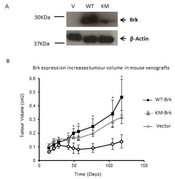
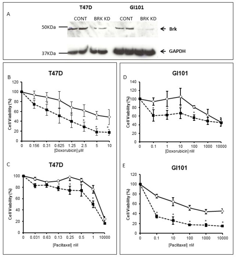
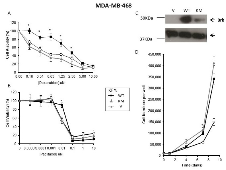
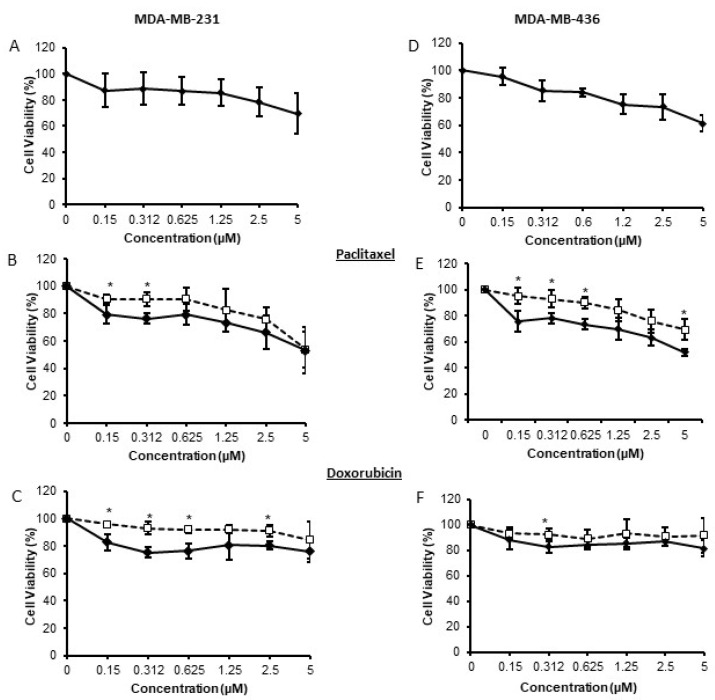
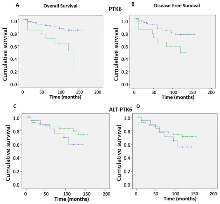
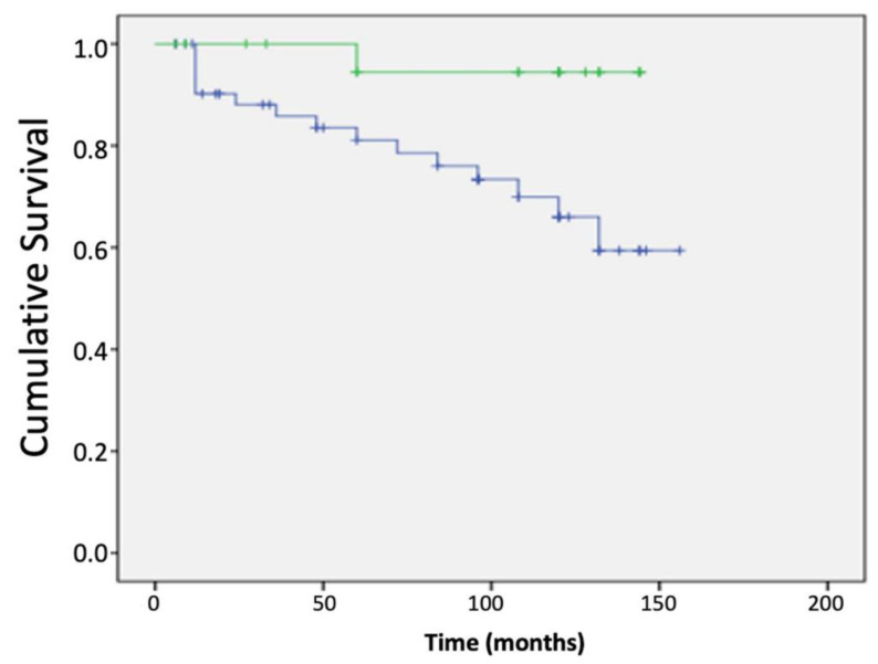
Breast tumour kinase (Brk/PTK6) is overexpressed in up to 86% of breast cancers and is associated with poorer patient outcomes. It is considered a potential therapeutic target in breast cancer, even though the full spectrum of its kinase activity is not known. This study investigated the role of the kinase domain in promoting tumour growth and its potential in sensitising triple negative breast cancer cells to standard of care chemotherapy. Triple negative human xenograft models revealed that both kinase-inactive and wild-type Brk promoted xenograft growth. Suppression of Brk activity in cells subsequently co-treated with the chemotherapy agents doxorubicin or paclitaxel resulted in an increased cell sensitivity to these agents. In triple negative breast cancer cell lines, the inhibition of Brk kinase activity augmented the effects of doxorubicin or paclitaxel. High expression of the alternatively spliced isoform, ALT-PTK6, resulted in improved patient outcomes. Our study is the first to show a role for kinase-inactive Brk in human breast tumour xenograft growth; therefore, it is unlikely that kinase inhibition of Brk, in isolation, would halt tumour growth in vivo. Breast cancer cell responses to chemotherapy in vitro were kinase-dependent, indicating that treatment with kinase inhibitors could be a fruitful avenue for combinatorial treatment. Of particular prognostic value is the ratio of ALT-PTK6:Brk expression in predicating patient outcomes.
Keywords: ALT-PTK6; Brk/PTK6; breast cancer; chemotherapy; kinase; prognosis.
Conflict of interest statement
The authors declare no conflict of interest.
Figures
Similar articles
-
Taxol Induces Brk-dependent Prosurvival Phenotypes in TNBC Cells through an AhR/GR/HIF-driven Signaling Axis.Mol Cancer Res. 2018 Nov;16(11):1761-1772. doi: 10.1158/1541-7786.MCR-18-0410. Epub 2018 Jul 10. Mol Cancer Res. 2018. PMID: 29991529 Free PMC article.
-
Breast Tumor Kinase (Brk/PTK6) Is Induced by HIF, Glucocorticoid Receptor, and PELP1-Mediated Stress Signaling in Triple-Negative Breast Cancer.Cancer Res. 2016 Mar 15;76(6):1653-63. doi: 10.1158/0008-5472.CAN-15-2510. Epub 2016 Jan 29. Cancer Res. 2016. PMID: 26825173 Free PMC article.
-
Mammary gland specific expression of Brk/PTK6 promotes delayed involution and tumor formation associated with activation of p38 MAPK.Breast Cancer Res. 2011 Sep 17;13(5):R89. doi: 10.1186/bcr2946. Breast Cancer Res. 2011. PMID: 21923922 Free PMC article.
-
Building a better understanding of the intracellular tyrosine kinase PTK6 - BRK by BRK.Biochim Biophys Acta. 2010 Aug;1806(1):66-73. doi: 10.1016/j.bbcan.2010.02.003. Epub 2010 Feb 26. Biochim Biophys Acta. 2010. PMID: 20193745 Free PMC article. Review.
-
Brk/PTK6 signaling in normal and cancer cell models.Curr Opin Pharmacol. 2010 Dec;10(6):662-9. doi: 10.1016/j.coph.2010.08.007. Epub 2010 Sep 9. Curr Opin Pharmacol. 2010. PMID: 20832360 Free PMC article. Review.
Cited by
-
Therapeutic Potential of Protein Tyrosine Kinase 6 in Colorectal Cancer.Cancers (Basel). 2023 Jul 21;15(14):3703. doi: 10.3390/cancers15143703. Cancers (Basel). 2023. PMID: 37509364 Free PMC article. Review.
-
Targeted Therapy and Mechanisms of Drug Resistance in Breast Cancer.Cancers (Basel). 2023 Feb 19;15(4):1320. doi: 10.3390/cancers15041320. Cancers (Basel). 2023. PMID: 36831661 Free PMC article. Review.
-
A PROTAC degrader suppresses oncogenic functions of PTK6, inducing apoptosis of breast cancer cells.Cell Chem Biol. 2025 Feb 20;32(2):255-266.e8. doi: 10.1016/j.chembiol.2024.10.008. Epub 2024 Nov 13. Cell Chem Biol. 2025. PMID: 39541980
References
Publication types
MeSH terms
Substances
LinkOut - more resources
Full Text Sources
