Review
. 2022 Feb 27;13(3):437.
doi: 10.3390/genes13030437.

Recombination Mediator Proteins: Misnomers That Are Key to Understanding the Genomic Instabilities in Cancer

Affiliations
Review

Recombination Mediator Proteins: Misnomers That Are Key to Understanding the Genomic Instabilities in Cancer

Justin Courcelle et al. Genes (Basel). .

Abstract

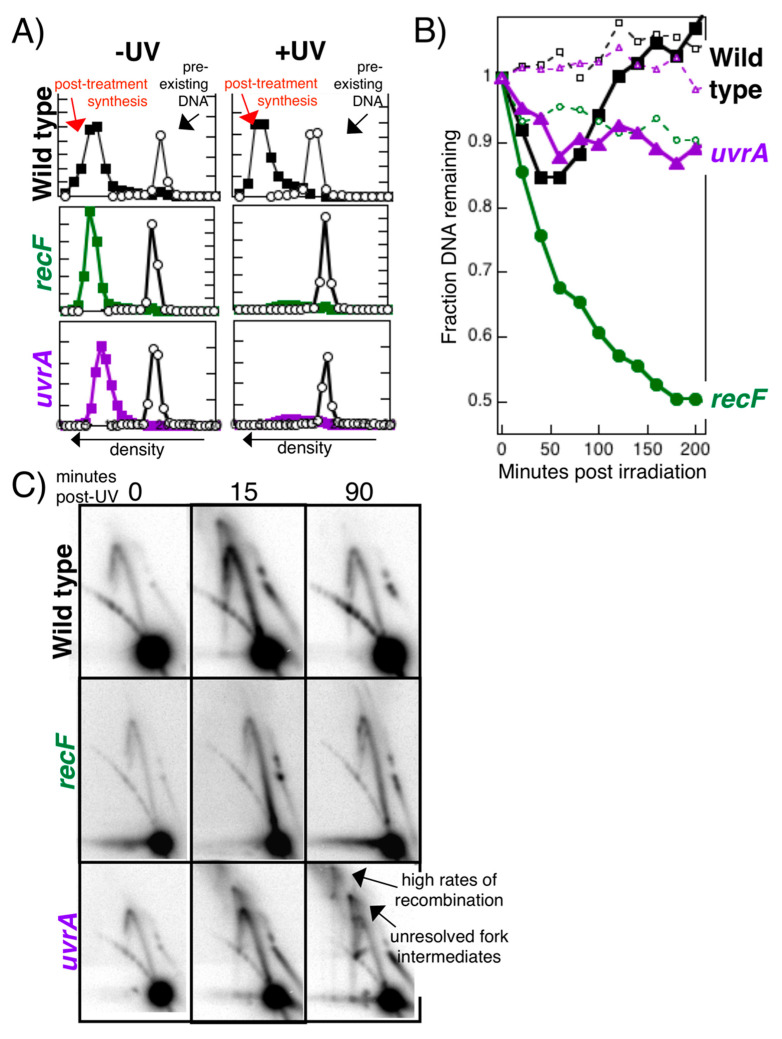
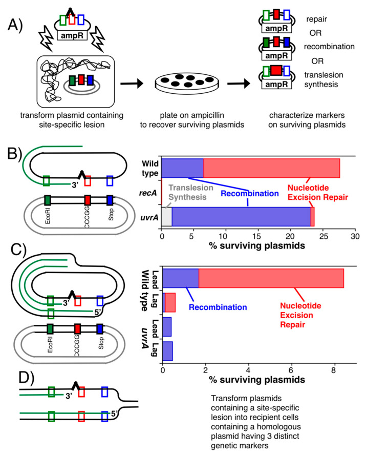
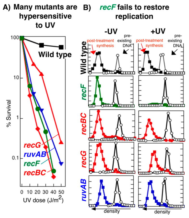
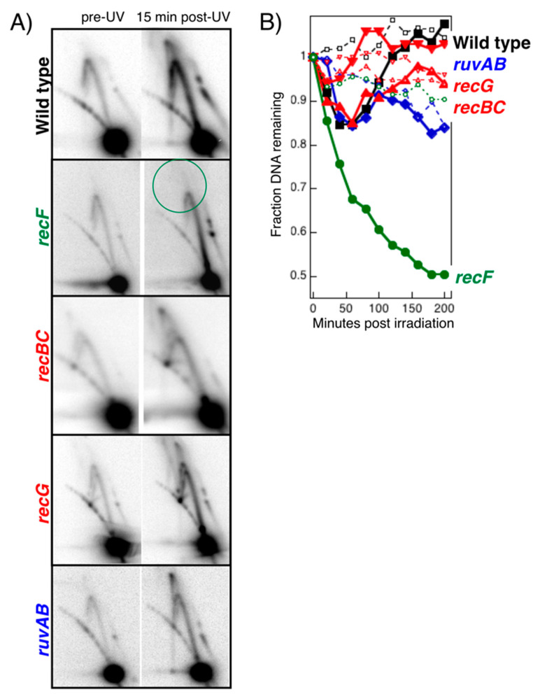
Recombination mediator proteins have come into focus as promising targets for cancer therapy, with synthetic lethal approaches now clinically validated by the efficacy of PARP inhibitors in treating BRCA2 cancers and RECQ inhibitors in treating cancers with microsatellite instabilities. Thus, understanding the cellular role of recombination mediators is critically important, both to improve current therapies and develop new ones that target these pathways. Our mechanistic understanding of BRCA2 and RECQ began in Escherichia coli. Here, we review the cellular roles of RecF and RecQ, often considered functional homologs of these proteins in bacteria. Although these proteins were originally isolated as genes that were required during replication in sexual cell cycles that produce recombinant products, we now know that their function is similarly required during replication in asexual or mitotic-like cell cycles, where recombination is detrimental and generally not observed. Cells mutated in these gene products are unable to protect and process replication forks blocked at DNA damage, resulting in high rates of cell lethality and recombination events that compromise genome integrity during replication.

Keywords: RecF; RecJ; RecO; RecQ; RecR; nucleotide excision repair; recombination; translesion synthesis.


Conflict of interest statement

The authors declare no conflict of interest.

Figures

Figure 1
Figure 1
Recombination frequency during nonsexual cell cycles correlates with genomic instability and cell death. (A) One form of recombination, sister chromatid exchanges, can be observed directly by growing cultured cells in 5-bromo-deoxyuridine for two generations before staining with Giemsa. In healthy cells, few exchanges are observed. By contrast, in cells lacking BLM, a RecQ homolog, recombination events are observed more frequently, and patients are predisposed to developing cancer. RecQ in E. coli is similarly required to suppress illegitimate recombination from occurring [17]. Photos generously provided by S. Wolff and A. A. Sandberg. (B) Experiment demonstrating that DNA damage-induced recombinational exchanges can be observed during replication in E. coli. By growing cells in different isotopic media, the DNA made before and after UV irradiation can be separated based upon their buoyant density in isopycnic alkali CsCl gradients. To test whether recombination can be induced by UV lesions, uvrA mutants defective in nucleotide excision repair were UV irradiated and allowed to recover for one hour. Unlike unirradiated uvrA mutants or irradiated wild-type cells, the DNA made in irradiated uvrA cultures contained more DNA at an intermediate density, demonstrating exchanges between the parental and daughter DNA strands are occurring in these cells (redrawn from [18] which reproduce results reported in [7]). (C) and (D) Although these exchanges were originally interpreted to represent a repair mechanism, the populations in which they were observed and characterized were rendered inviable by the UV treatment.
Figure 2
Figure 2
Substrates generated during replication on UV-damaged templates. Based upon our current understanding, lesions in the leading-strand template disrupt replisome progression. In contrast, lesions in the lagging-strand template produce gaps but do not disrupt replication.
Figure 3
Figure 3
Model of how the RecF pathway restores replication following disruption. ^, pyrimidine dimer; purple subunits, the tau complex, DNA polymerase III, and processivity factors of the replisome; green subunits, helicase-primase; Q, RecQ; J, RecJ: F, RecF; O, RecO; R, RecR; A, RecA. (AH) Different RecF pathway gene products.
Figure 4
Figure 4
RecF is required to maintain replication forks disrupted by DNA damage. (A) recF mutants fail to restore replication following disruption by UV-induced damage. The amount of replication occurring within 1 h after UV irradiation with 25 J/m2 was analyzed by alkaline CsCl density gradients. Cells prelabeled with [14C]thymine were irradiated or mock-irradiated, filtered, and allowed to recover for 1 h in media containing [3H]5-bromodeoxyuridine to density label DNA made during this period. Circles, 14C prelabeled DNA; squares, 3H replicated DNA. (B) recF mutants are unable to maintain the replication fork processing observed by two-dimensional gel electrophoresis. The migration pattern in 2D agarose gels for linearized pBR322 plasmid after UV treatment. Nonreplicating plasmids run as a linear 4.4 kb fragment. Normal replicating plasmids form Y-shaped structures and migrate more slowly due to their increased size and nonlinear shape, moving as an arc that extends from the linear fragment. Replication intermediates observed after UV damage form double-Y or X-shaped structures that migrate in the cone region. Whereas processing intermediates transiently accumulate in wild-type cells after UV irradiation, no intermediates are observed in recF mutants. (C) The nascent DNA undergoes excessive degradation in recF mutants. [3H]thymidine is added to [14C]thymine-prelabeled cells for 10 s immediately before the cells were filtered and irradiated with 25 J/m2 in nonradioactive medium. The fraction of the radioactivity remaining in the DNA is plotted against time. Loss of 14C genomic DNA (open symbols) can be compared to the loss of the 3H DNA synthesized at the growing fork just prior to irradiation (filled symbols). Squares, wild type; circles, recF. Compiled and redrawn from data reported in [18,53].
Figure 5
Figure 5
The RecQ helicase and RecJ nuclease degrade the nascent lagging strand of disrupted replication forks. (A) The nascent DNA degradation at disrupted replication forks does not occur in the absence of either RecQ or RecJ. Degradation of nascent replication fork DNA was measured as described in Figure 4. In recF or recR mutants, excessive degradation of the nascent DNA occurs after UV. However, this degradation does not occur in the absence of RecJ or RecQ. Filled symbols, nascent DNA; open symbols, total genomic DNA. (B) Inactivation of the RecJ nuclease or RecQ helicase restores the processing intermediates to recF mutants as observed in 2D agarose gels. Cells containing the plasmid pBR322 were UV irradiated and analyzed by 2D agarose gel analysis as in Figure 4. (C) RecJ preferentially degrades the nascent lagging strand of disrupted replication forks. Nascent DNA from irradiated cultures was isolated by pulse-labelling cultures with [3H]5-bromodeoxyuridine, followed by separation in alkali CsCl gradients. The leading- and lagging-strand nascent DNA remaining at each time point were quantified by using strand-specific probes of the lacZ gene. The ratio of the lagging strand signal to the leading strand signal at each time point is plotted. Redrawn from data reported in [51,53].
Figure 6
Figure 6
Model for how RecQ and RecJ suppress rearrangements and mutations. (A) RecJ-RecQ-mediated degradation of the nascent lagging strand at arrested replication forks (i) restores the lesion-containing region to a double-stranded form that can be repaired (ii) and creates a large substrate (region of single-stranded DNA with homologous duplex DNA) at the fork that RecF-O-R and RecA can bind and stabilize (iii) so that replication can resume (iv). (B) In the absence of RecJ-RecQ degradation, the region containing the lesion remains in single-stranded form, preventing its repair, and delaying the recovery until translesion synthesis can occur (v). Additionally, in the absence of RecJ-RecQ processing, RecF-O-R and RecA are unable to bind, leading to recombination at the disrupted site and compromising viability if translesion synthesis cannot occur (vi).
Figure 7
Figure 7
In the absence of RecJ processing, repair cannot occur and replication recovery and cell survival become dependent on translesion synthesis. (A) In the absence of RecJ, the recovery of replication is delayed and becomes entirely dependent on translesion synthesis by PolV, encoded by umuC. To measure the time at which replication resumes, [3H]thymidine was added to cultures for 2 min at the indicated times following either 27 J/m2 UV irradiation (filled symbols) or mock irradiation (open symbols) at time 0. The relative amount of DNA synthesis/2 min, 3H (squares), is plotted. The time replication resumes is indicated by the red line on each graph. (B) In the absence of RecJ, survival becomes dependent on translesion synthesis by Pol V. The relative survival of each culture following UV irradiation with the indicated dose is shown. A fresh overnight culture was evenly applied onto a Luria-Bertani medium plate with a cotton swab. The plate was covered, placed under a UV lamp, and the cover was progressively retracted following 20 J/m2 exposures. Redrawn from data reported in [55,56].
Figure 8
Figure 8
Survival following UV irradiation requires both rec and uvr function. The number of UV-induced lesions that wild-type, uvrA, recA and recA uvrA cells can survive is plotted. Composed from data reported in [49].
Figure 9
Figure 9
Restoring replication requires both nucleotide excision repair and the RecF pathway. (A) Replication fails to resume in the absence of either RecF or nucleotide excision repair. The amount of replication occurring within 1 h after UV irradiation with 25 J/m2 was analyzed by alkaline CsCl density gradients as described in Figure 4. Circles, 14C-prelabeled DNA; squares, 3H-replicated DNA. (B) Unlike in recF mutants, the replication fork remains protected in nucleotide excision repair mutants. The nascent DNA degradation occurring after UV irradiation with 25 J/m2 was measured as described in Figure 4. Loss of 14C genomic DNA (open symbols) can be compared to the loss of the 3H DNA synthesized at the growing fork just prior to irradiation (filled symbols). Squares, wild type; circles, recF; triangles, uvrA. (C) In the absence of nucleotide excision repair, the intermediates at disrupted replication forks are not resolved and aberrant, higher order recombination intermediates accumulate. The migration pattern of replicating plasmids in each strain was analyzed by 2D agarose gel analysis as described in Figure 4. Processing intermediates transiently accumulate and then resolve in wild-type cells. No intermediates are observed in recF mutants. In uvrA mutants, these intermediates persist and go on to form higher-order, recombination intermediates. Redrawn from data reported in [18,53].
Figure 10
Figure 10
Nucleotide excision repair occurs at disrupted replication forks. (A) Schematic to measure lesion processing on replicating plasmids. Plasmids containing a site-specific AAF adduct were transformed into cells containing a second endogenous, homologous plasmid with three closely spaced, genetic markers. The markers are designed to allow one to determine whether nucleotide excision repair (surviving plasmids only contained the marker at the lesion site), recombination (surviving plasmids contained multiple genetic markers), or translesion synthesis (contained a base-pair insertion at the middle marker) occurred on the surviving plasmids that are recovered. (B) Nucleotide excision repair events predominate at disrupted replication forks. When single-stranded plasmids were used in this assay, nucleotide excision repair predominated over recombination events. Few plasmids were recovered in recA mutants. In the absence of nucleotide excision repair, survival was reduced and recombination events predominated. (C) Nucleotide excision repair occurs when the lesion is in the leading strand, which disrupts replication, but not the lagging strand, which does not disrupt replication. The same assay is repeated as in (B), except transforming a double-stranded plasmid containing an AAF adduct in either the leading- or lagging-strand template. In wild-type cells, nucleotide excision repair events predominated on the recovered plasmids when the lesion was in the leading strand. When the lesion was in the lagging strand the frequency of repair and plasmids recovered was severely reduced. In uvrA mutants, the frequency of plasmids recovered was severely reduced irrespective of whether the lesion was in the leading- or lagging-strand template and recombination events predominated. (D) To be scored in this assay, repair events were required to undergo strand exchange and utilize an intermolecular template for repair. In vivo, the template utilized for repair would be intramolecular, as shown, allowing the reaction to occur with far, far greater frequency and efficiency. Composed and drawn from data reported in [83,84].
Figure 11
Figure 11
Restoring replication after disruption is specific to the recF pathway. (A) recF, recBC, ruvAB, and recG mutants all exhibit high levels of cell lethality following UV-induced DNA damage. The survival of UV-irradiated wild-type, recF, recBC, ruvAB, and recG cells at the doses indicated is plotted. (B) Yet only recF mutants fail to restore replication after UV-induced damage. The amount of DNA synthesized during a 1 h period in UV-irradiated (25 J/m2) or mock-irradiated cultures was determined by density labeling the DNA with 5-bromodeoxyuridine and subsequent isolation in isopycnic alkali CsCl gradients. Open circles, DNA synthesized before treatment (14C); filled squares, DNA synthesized following treatment (3H). The range of 3H and 14C axes was kept constant for each strain. Redrawn from data presented in [91,164,165].
Figure 12
Figure 12
Processing of disrupted replication forks is specific to the RecF pathway. (A) The absence of processing intermediates seen at disrupted replication fork is specific to recF mutants. DNA from cells containing the plasmid pBR322 were purified, digested with Pvu II, and analyzed by 2D agarose gels immediately before and 15 min after UV irradiation with 50 J/m2. Cone region intermediates resembling those in wild-type cells appear in recBC, recG, and ruvAB mutants, but are missing in recF mutants. (B) The inability to protect the nascent DNA from degradation is specific to the RecF pathway. A 10 s pulse of [3H]thymidine is added to [14C]thymine-prelabeled cells immediately before the cells are filtered and irradiated with 25 J/m2 in nonradioactive medium. The loss (or degradation) of 14C genomic DNA (open symbols) can be compared with the loss of the 3H DNA synthesized at the growing fork just before irradiation (filled symbols). Squares, wild type; circles, recF; diamonds, recBC; triangles, recG; inverted triangles, ruvAB. Data adapted from [53,80,91].

Similar articles

Cited by

References

    1. Lederberg J., Tatum E.L. Sex in Bacteria: Genetic Studies, 1945–1952. Science. 1953;118:169–175. doi: 10.1126/science.118.3059.169. - DOI - PubMed
    1. Clark A.J., Margulies A.D. Isolation and characterization of recombination-deficient mutants of Escherichia coli K12. Proc. Natl. Acad. Sci. USA. 1965;53:451–459. doi: 10.1073/pnas.53.2.451. - DOI - PMC - PubMed
    1. Clark A.J. The beginning of a genetic analysis of recombination proficiency. J. Cell. Physiol. 1967;70:165–180. doi: 10.1002/jcp.1040700412. - DOI - PubMed
    1. Horii Z., Clark A.J. Genetic analysis of the recF pathway to genetic recombination in Escherichia coli K12: Isolation and characterization of mutants. J. Mol. Biol. 1973;80:327–344. doi: 10.1016/0022-2836(73)90176-9. - DOI - PubMed
    1. Howard-Flanders P., Theriot L. Mutants of Escherichia coli K-12 defective in DNA repair and in genetic recombination. Genetics. 1966;53:1137–1150. doi: 10.1093/genetics/53.6.1137. - DOI - PMC - PubMed

Publication types