. 2022 Feb 27;13(3):436.
doi: 10.3390/genes13030436.

The Effect of Dicer Knockout on RNA Interference Using Various Dicer Substrate Small Interfering RNA (DsiRNA) Structures

Affiliations

The Effect of Dicer Knockout on RNA Interference Using Various Dicer Substrate Small Interfering RNA (DsiRNA) Structures

Min-Sun Song et al. Genes (Basel). .

Abstract

Small interfering RNAs (siRNAs) are artificial molecules used to silence genes of interest through the RNA interference (RNAi) pathway, mediated by the endoribonuclease Dicer. Dicer-substrate small interfering RNAs (DsiRNAs) are an alternative to conventional 21-mer siRNAs, with an increased effectiveness of up to 100-fold compared to traditional 21-mer designs. DsiRNAs have a novel asymmetric design that allows them to be processed by Dicer into the desired conventional siRNAs. DsiRNAs are a useful tool for sequence-specific gene silencing, but the molecular mechanism underlying their increased efficacy is not precisely understood. In this study, to gain a deeper understanding of Dicer function in DsiRNAs, we designed nicked DsiRNAs with and without tetra-loops to target a specific mRNA sequence, established a Dicer knockout in the HCT116 cell line, and analyzed the efficacy of various DsiRNAs on RNAi-mediated gene silencing activity. The gene silencing activity of all DsiRNAs was reduced in Dicer knockout cells. We demonstrated that tetra-looped DsiRNAs exhibited increased efficacy for gene silencing, which was mediated by Dicer protein. Thus, this study improves our understanding of Dicer function, a key component of RNAi silencing, which will inform RNAi research and applications.

Keywords: CRISPR system; RNA biogenesis; RNA interference (RNAi); dicer; dicer knockout; dicer-substrate siRNA (DsiRNA); tetra-loop.


Conflict of interest statement

The authors declare no conflict of interest.

Figures

Figure 1
Figure 1
Dicer mutation using the CRISPR/Cas9 system. (A) Domain structure of Dicer protein. The small blue rectangle underneath indicates the region corresponding to the gDNA sequence targeted by the CRISPR/Cas9 system. (B) Sequence of DICER gDNA. The two sites with blue letters show the sequence targeted by double-nickase CRISPR. Green and red arrows indicate primers for detecting DICER gDNA.
Figure 2
Figure 2
A representative sequence of the human DICER locus targeted by Cas9. (A) Schematic illustrating DNA double-nick approach using a pair of sgRNAs to guide Cas9 nickase. Cas9 can cleave only the strand complementary to the sgRNA (blue letters). A pair of sgRNA-Cas9 can cleave Dicer gDNA. Expected Cas9 cleavage sites are marked as red arrows. sgRNA offset is characterized as the distance between the PAM sequence (brown letters) and the 5′-ends of the guide sequence of a given sgRNA pair. (B) Representative sequences of the gDNA in the helicase domain of human DICER targeted by sgRNA. The PAM area is represented by a dotted red underline (upper) or red box (bottom). The dotted vertical line is the expected cleavage site. H2-2 clone sequences were compared with the human DICER gDNA sequence (XP_016876610:NCBI reference sequence). The negative control is the gDNA sequence from parental WT HCT116.
Figure 3
Figure 3
Analysis of Dicer protein and mRNA in HCT116 and Dicer knockout H2-2 cells. (A) Western blot experiments to confirm gene disruption in H2-2 Dicer knockout cells compared to WT HCT116 cells. The Dicer protein is 217 kDa in humans. Two anti-Dicer antibodies (Ab14601 and Ab13502) were used to detect the Dicer protein. Tubulin was used as a loading control. (B) Amino acid sequence analysis of the mutated area from H2-2 and human Dicer. The CRISPR/Cas9 system made nine nucleotide deletions and twenty-one nucleotide substitutions (Supplementary Figure S2A). The Dicer sequence from H2-2 cells shows a different protein-peptide compared with human Dicer (NP_001182502.1: NCBI reference number). (C) Human endoribonuclease Dicer mRNA sequence (1 to 300 nt: NM_001195573.1: NCBI reference sequence). Red arrows show the forward and reverse primers used to detect Dicer mRNA by PCR. The CRISPR/Cas9 system mutated the sequence indicated with blue underline. Yellow slashes indicate the codon of amino acid. (D) RNA was extracted from HCT116 and H2-2 cells and subjected to reverse transcription either with or without (w/o RT) the RT enzyme. cDNA was amplified by PCR with a Dicer-targeted primer (red arrow in (C)). The gray arrow is the DNA band of 100 bp in size. The last lane in the gel lane loads the low molecular weight DNA ladder (New England BioLabs Inc.: catalog number N3233). Black arrow is size of 50 bp.
Figure 4
Figure 4
Structure of hnRNPH1-targeted DsiRNAs and tetra-looped DsiRNAs. We designed two DsiRNAs targeting hnRNPH1, DsiRNA I (DI) and DsiRNA II (DII), then added a 5′-GAAA-3′ tetra-loop (TL) into each DsiRNA and synthesized different stem structures for each, one with a GC-rich stem (TL_DI and TL_DII) and another with the original stem (TL_DI_O and TL_DII_O), which corresponds to the hnRNPH1 target gene. TL_DI has a 21-mer sense strand and a 38-mer antisense strand, with a GC-rich stem and 5′-GAAA-3′ tetra-loop (represented with blue circles). TL_DI_O has the same sequence as DI, with a 5′-GAAA-3′ tetra-loop sequence (blue circles). TL_DII has a 22-mer antisense strand and 36-mer sense strand, with a GC-rich stem and 5′-GAAA-3′ tetra-loop (blue circles). TL_DII_O has the same sequence as DII, with a 5′-GAAA-3′ tetra-loop sequence (blue circles). The detailed sequences are listed in Table 1. Ribonucleotides are shown in upper case and deoxyribonucleotides as dN. The purple underlined sequence shows the active strand. The blue arrow indicates the nick between ribonucleotides.
Figure 5
Figure 5
Surveyor assay comparing the efficiency of Cas9-mediated cleavage by double-nickase sgRNA in the human DICER locus. DNA duplex formation and treatment with a nuclease. SURVEYOR assay gel showing a comparable modification of control G/C, which is 633 base pair (bp) Control DNA with a point mutation (Supplementary Figure S1B) bearing 416 bp and 217 bp. Homoduplex without mismatch did not cleave the nuclease, but heteroduplex (Dicer H2-1, H2-2, and H2-3) shows the cleavage band. We ran the gel with short (A) and long (B) running times. Arrowheads indicate cleavage products.
Figure 6
Figure 6
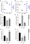
DsiRNA efficacy in HCT116 cells. Humanized Renilla luciferase was cloned in either the sense or antisense orientation into hnRNPH1 transcripts to act as a reporter. Relative expression of Renilla luciferase was determined by dual-luciferase assay against an internal control of firefly luciferase; Renilla activities were normalized to those of firefly and arbitrarily set at 100. Data represent the mean ± S.D. from three independent experiments (Student’s t-test ** p < 0.01, *** p < 0.001, and **** p < 0.0001). We transfected the dual-luciferase reporter (S reporter targeting the sense strand of DsiRNAs, AS reporter targeting the antisense strand of DsiRNAs) and variant DsiRNAs into HCT116 cells. The DsiRNAs represented are: (A). DsiRNA I, (B). DsiRNA II, (C): DsiRNA I_S, (D): DsiRNA II_S, (E): DsiRNA I_AS, and (F): DsiRNA II_AS.
Figure 7
Figure 7
Comparison of DsiRNA efficacy in HCT116 and Dicer knockout H2-2 cells. The detection method used was the same as in Figure 6. We transfected the dual-luciferase reporter and DsiRNAs into HCT116 (black) and Dicer knockout H2-2 (red) cells. We determined the fold decrease of gene silencing efficiency in H2-2 by calculating the activity of H2-2 divided by HCT116 (green bar), as represented on the right y-axis. Graphs show the gene silencing activity of sense (S) strands (AF) of (A): DI, (B): DI_TL, (C): DI_TL_O, (D): DII, (E): DII_TL, and (F): DII_TL_O, and antisense (AS) strands (GL) of (G): DI, (H): DI_TL, (I): DI_TL_O, (J): DII, (K): DII_TL, and (L): DII_TL_O. Data represent the mean ± S.D. of three independent experiments (Student’s t-test * p < 0.05, ** p < 0.01, *** p < 0.001, and **** p < 0.0001).
Figure 7
Figure 7
Comparison of DsiRNA efficacy in HCT116 and Dicer knockout H2-2 cells. The detection method used was the same as in Figure 6. We transfected the dual-luciferase reporter and DsiRNAs into HCT116 (black) and Dicer knockout H2-2 (red) cells. We determined the fold decrease of gene silencing efficiency in H2-2 by calculating the activity of H2-2 divided by HCT116 (green bar), as represented on the right y-axis. Graphs show the gene silencing activity of sense (S) strands (AF) of (A): DI, (B): DI_TL, (C): DI_TL_O, (D): DII, (E): DII_TL, and (F): DII_TL_O, and antisense (AS) strands (GL) of (G): DI, (H): DI_TL, (I): DI_TL_O, (J): DII, (K): DII_TL, and (L): DII_TL_O. Data represent the mean ± S.D. of three independent experiments (Student’s t-test * p < 0.05, ** p < 0.01, *** p < 0.001, and **** p < 0.0001).

References

    1. Cummings J.C., Zhang H., Jakymiw A. Peptide carriers to the rescue: Overcoming the barriers to siRNA delivery for cancer treatment. Transl. Res. J. Lab. Clin. Med. 2019;214:92–104. doi: 10.1016/j.trsl.2019.07.010. - DOI - PMC - PubMed
    1. Adams D., Gonzalez-Duarte A., O’Riordan W.D., Yang C.C., Ueda M., Kristen A.V., Tournev I., Schmidt H.H., Coelho T., Berk J.L., et al. Patisiran, an RNAi Therapeutic, for Hereditary Transthyretin Amyloidosis. N. Engl. J. Med. 2018;379:11–21. doi: 10.1056/NEJMoa1716153. - DOI - PubMed
    1. Raja M.A.G., Katas H., Amjad M.W. Design, mechanism, delivery and therapeutics of canonical and Dicer-substrate siRNA. Asian J. Pharm. Sci. 2019;14:497–510. doi: 10.1016/j.ajps.2018.12.005. - DOI - PMC - PubMed
    1. Soukup G.A. Encyclopedia of Life Sciences. Wiley & Sons; Hoboken, NJ, USA: 2003. Nucleic acids: General properties. - DOI
    1. Chen Y., Varani G. RNA Structure. In: eLS , editor. Encyclopedia of Life Sciences. Wiley & Sons; Hoboken, NJ, USA: 2010.

Publication types

MeSH terms

Substances

LinkOut - more resources