Case Reports
. 2022 Mar 8;13(3):478.
doi: 10.3390/genes13030478.

First Description of Inheritance of a Postzygotic OPA1 Mosaic Variant

Affiliations
Case Reports

First Description of Inheritance of a Postzygotic OPA1 Mosaic Variant

Svenja Alter et al. Genes (Basel). .

Abstract

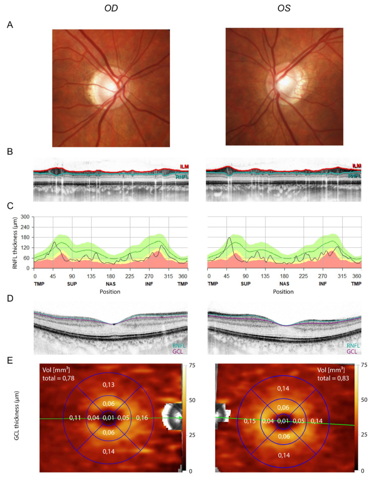
Optic atrophy 1 (MIM #165500) is caused by pathogenic variants in the gene OPA1 (OPA1 MITOCHONDRIAL DYNAMIN-LIKE GTPase, MIM *605290) and is inherited in an autosomal dominant manner. We describe a 6-year-old male patient with severe early onset manifestation of optic atrophy, whose parents are subjectively asymptomatic. OPA1-sequence analysis revealed the heterozygous missense variant NM_015560.3:c.806C>T, p.(Ser269Phe) in the patient. Segregation analysis of the parents showed that the mother carried a low-grade postzygotic mosaic of this variant, which apparently also involves germline cells. In line with this, ophthalmological investigation of the mother showed subclinical manifestation of optic atrophy 1. This is the first report of an OPA1 postzygotic mosaic that was inherited to offspring.

Keywords: ADOA; OPA1; optic atrophy; postzygotic mosaic.


Conflict of interest statement

The authors declare no conflict of interest.

Figures

Figure 1
Figure 1
Severe optic atrophy in the son. (A) Fundus images showing temporally accentuated optic pallor. (B) Circumpapillary OCT scan (ILM in red and the border between GCL and RNFL in turquoise). Note the thinning of the RNFL. (C) Peripapillary RNFL thickness in all sectors, showing severe circular optic atrophy. (D) Goldmann perimetry showing a relatively central scotoma. Isopters: red = I3; dark blue = I4; green = III4. Note that isopter I1 was not seen. OD = right eye, OS = left eye, OCT = optical coherence tomography, ILM = internal limiting membrane, GCL = ganglion cell layer.
Figure 2
Figure 2
Next generation sequencing results. The variant NM_015560.3:c.806C>T, p.(Ser269Phe) in exon 8 of the OPA1 gene was detected in a heterozygous state in DNA isolated from peripheral blood of the index patient (VAF = 54.9%). In the mother, the variant was detected with a mosaic of 15.3% in peripheral blood, 18.4% in oral mucosa and 10.0% in skin. VAF = variant allele frequency.
Figure 3
Figure 3
Mild optic atrophy in the mother. (A) Fundus images showing mild myopic papillary changes. (B) Circumpapillary OCT scan (ILM in red, border between GCL and RNFL in turquoise). (C) Macular OCT scan (border between inner nuclear layer and GCL in purple, border between GCL and RNFL in turquoise). (D,E) GCL volume quantification from macular OCT. Note the mild reduction in GCL volume (mean in the respective age group = 1.12 mm³, 5th percentile = 0.99 mm³, 95th percentile = 1.25 mm³; reference data from Meyer et al. (2021) [25]).

References

    1. Ferré M., Bonneau D., Milea D., Chevrollier A., Verny C., Dollfus H., Ayuso C., Defoort S., Vignal C., Zanlonghi X., et al. Molecular Screening of 980 Cases of Suspected Hereditary Optic Neuropathy with a Report on 77 Novel OPA1 Mutations. Hum. Mutat. 2009;30:E692–E705. doi: 10.1002/humu.21025. - DOI - PubMed
    1. Newman N.J. Hereditary Optic Neuropathies: From the Mitochondria to the Optic Nerve. Am. J. Ophthalmol. 2005;140:517.e1–517.e9. doi: 10.1016/j.ajo.2005.03.017. - DOI - PubMed
    1. Cipolat S., Martins de Brito O., Dal Zilio B., Scorrano L. OPA1 Requires Mitofusin 1 to Promote Mitochondrial Fusion. Proc. Natl. Acad. Sci. USA. 2004;101:15927–15932. doi: 10.1073/pnas.0407043101. - DOI - PMC - PubMed
    1. Frezza C., Cipolat S., Martins de Brito O., Micaroni M., Beznoussenko G.V., Rudka T., Bartoli D., Polishuck R.S., Danial N.N., De Strooper B., et al. OPA1 Controls Apoptotic Cristae Remodeling Independently from Mitochondrial Fusion. Cell. 2006;126:177–189. doi: 10.1016/j.cell.2006.06.025. - DOI - PubMed
    1. Yu-Wai-Man P., Trenell M.I., Hollingsworth K.G., Griffiths P.G., Chinnery P.F. OPA1 Mutations Impair Mitochondrial Function in Both Pure and Complicated Dominant Optic Atrophy. Brain. 2011;134:e164. doi: 10.1093/brain/awq288. - DOI - PMC - PubMed

Publication types

LinkOut - more resources