

Trauma of Peripheral Innervation Impairs Content of Epidermal Langerhans Cells
- PMID: 35328120
- PMCID: PMC8947052
- DOI: 10.3390/diagnostics12030567
Trauma of Peripheral Innervation Impairs Content of Epidermal Langerhans Cells
Abstract
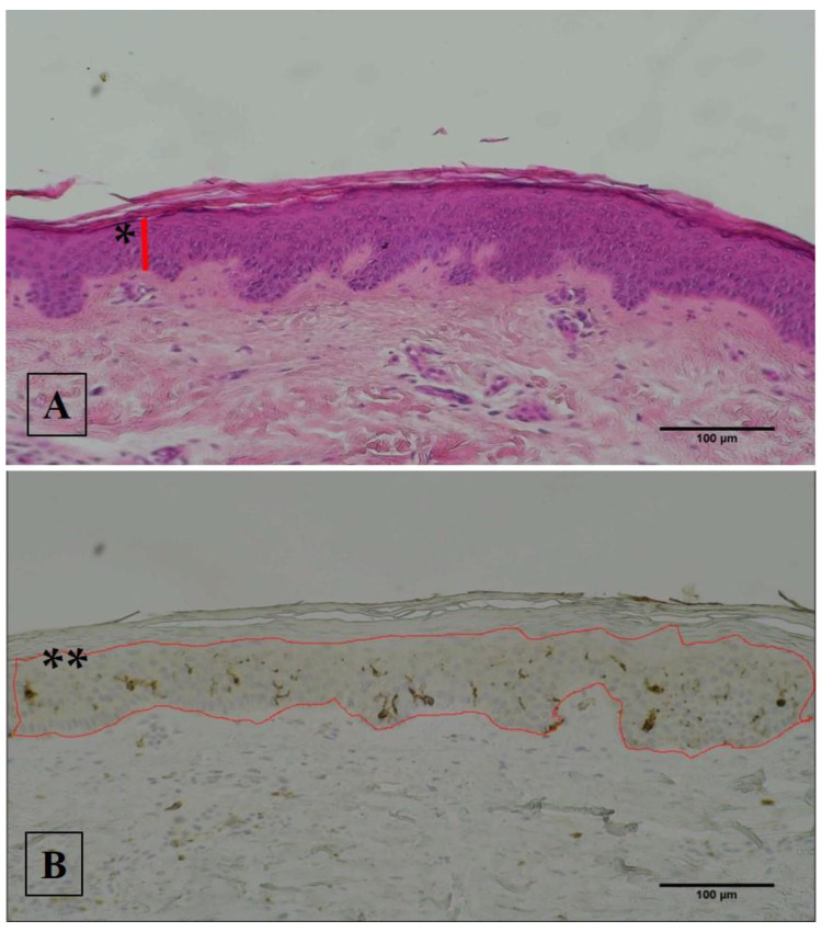
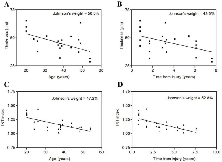
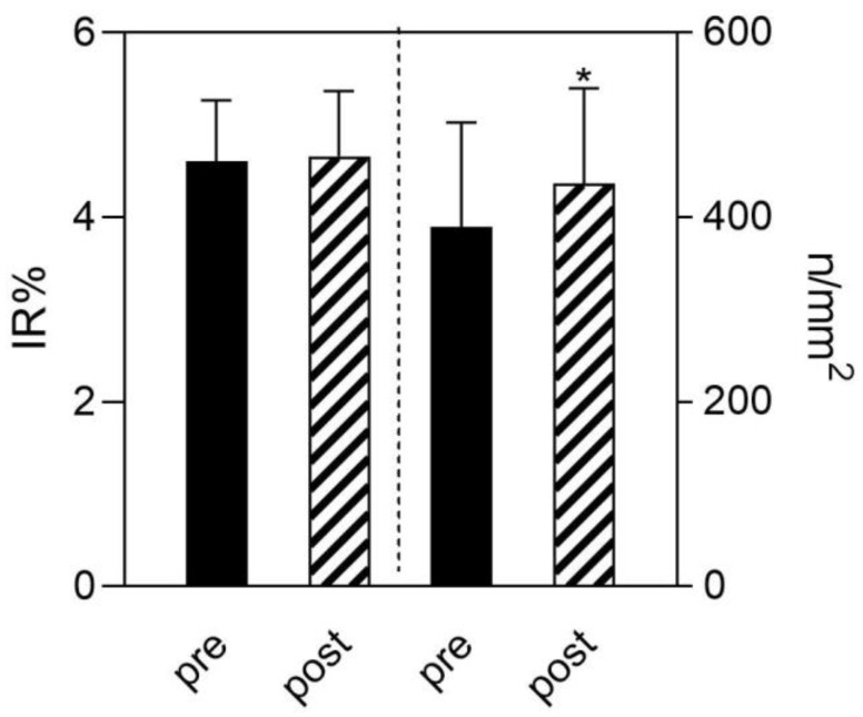
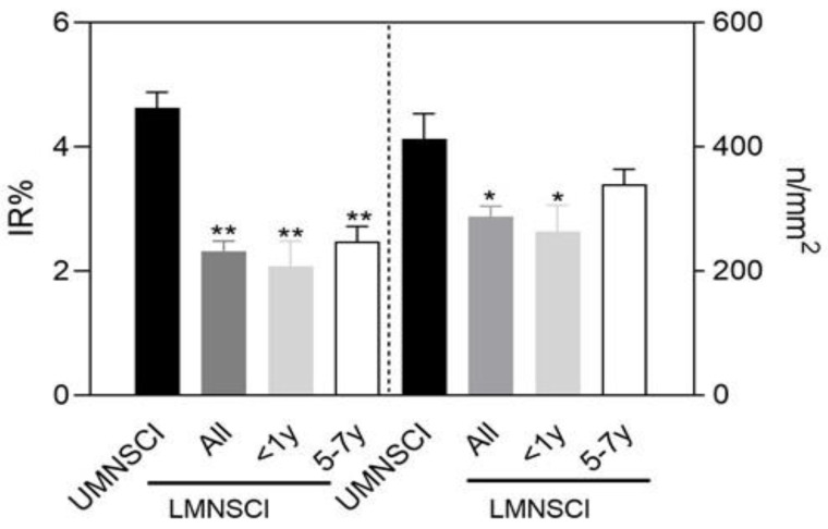
Langerhans cells represent the first immune cells that sense the entry of external molecules and microorganisms at the epithelial level in the skin. In this pilot case-study, we evaluated Langerhans cells density and progression of epidermal atrophy in permanent spinal cord injury (SCI) patients suffering with either lower motor neuron lesions (LMNSCI) or upper motor neuron lesions (UMNSCI), both submitted to surface electrical stimulation. Skin biopsies harvested from both legs were analyzed before and after 2 years of home-based Functional Electrical Stimulation for denervated degenerating muscles (DDM) delivered at home (h-bFES) by large anatomically shaped surface electrodes placed on the skin of the anterior thigh in the cases of LMNSCI patients or by neuromuscular electrical stimulation (NMES) for innervated muscles in the cases of UMNSCI persons. Using quantitative histology, we analyzed epidermal thickness and flattening and content of Langerhans cells. Linear regression analyses show that epidermal atrophy worsens with increasing years of LMNSCI and that 2 years of skin electrostimulation reverses skin changes, producing a significant recovery of epidermis thickness, but not changes in Langerhans cells density. In UMNSCI, we did not observe any statistically significant changes of the epidermis and of its content of Langerhans cells, but while the epidermal thickness is similar to that of first year-LMNSCI, the content of Langerhans cells is almost twice, suggesting that the LMNSCI induces an early decrease of immunoprotection that lasts at least 10 years. All together, these are original clinically relevant results suggesting a possible immuno-repression in epidermis of the permanently denervated patients.
Keywords: Langerhans cells; electrical stimulation; epidermis atrophy; lower or upper motor neuron injury; skin biopsy; spinal cord injury.
Conflict of interest statement
The authors declare no conflict of interest.
Figures
Similar articles
-
Two-years of home based functional electrical stimulation recovers epidermis from atrophy and flattening after years of complete Conus-Cauda Syndrome.Medicine (Baltimore). 2019 Dec;98(52):e18509. doi: 10.1097/MD.0000000000018509. Medicine (Baltimore). 2019. PMID: 31876739 Free PMC article. Clinical Trial.
-
Dermal papillae flattening of thigh skin in Conus Cauda Syndrome.Eur J Transl Myol. 2018 Dec 13;28(4):7914. doi: 10.4081/ejtm.2018.7914. eCollection 2018 Nov 2. Eur J Transl Myol. 2018. PMID: 30662702 Free PMC article.
-
In complete SCI patients, long-term functional electrical stimulation of permanent denervated muscles increases epidermis thickness.Neurol Res. 2018 Apr;40(4):277-282. doi: 10.1080/01616412.2018.1436877. Epub 2018 Feb 15. Neurol Res. 2018. PMID: 29447083
-
Atrophy, ultra-structural disorders, severe atrophy and degeneration of denervated human muscle in SCI and Aging. Implications for their recovery by Functional Electrical Stimulation, updated 2017.Neurol Res. 2017 Jul;39(7):660-666. doi: 10.1080/01616412.2017.1314906. Epub 2017 Apr 13. Neurol Res. 2017. PMID: 28403681 Review.
-
Electrical stimulation and denervated muscles after spinal cord injury.Neural Regen Res. 2020 Aug;15(8):1397-1407. doi: 10.4103/1673-5374.274326. Neural Regen Res. 2020. PMID: 31997798 Free PMC article. Review.
Cited by
-
Langerhans cells: Central players in the pathophysiology of atopic dermatitis.J Eur Acad Dermatol Venereol. 2025 Feb;39(2):278-289. doi: 10.1111/jdv.20291. Epub 2024 Aug 19. J Eur Acad Dermatol Venereol. 2025. PMID: 39157943 Free PMC article. Review.
-
Abstracts of the 2023 Padua Days of Muscle and Mobility Medicine (2023Pdm3) to be held March 29 - April 1 at the Galileian Academy of Padua and at the Petrarca Hotel, Thermae of Euganean Hills, Padua, Italy.Eur J Transl Myol. 2023 Feb 10;33(1):11247. doi: 10.4081/ejtm.2023.11247. Online ahead of print. Eur J Transl Myol. 2023. PMID: 36786151 Free PMC article.
-
2023 Padua Days of Muscle and Mobility Medicine: post-meeting Book of Abstracts.Eur J Transl Myol. 2023 Apr 27;33(2):11427. doi: 10.4081/ejtm.2023.11427. Eur J Transl Myol. 2023. PMID: 37114363 Free PMC article.
References
-
- Kern H., Hofer C., Loefler S., Zampieri S., Gargiulo P., Baba A., Marcante A., Piccione F., Pond A., Carraro U. Atrophy, ultra-structural disorders, severe atrophy and degeneration of denervated human muscle in SCI and Aging. Implications for their recovery by Functional Electrical Stimulation, updated 2017. Neurol. Res. 2017;39:660–666. doi: 10.1080/01616412.2017.1314906. - DOI - PubMed
LinkOut - more resources
Full Text Sources
