

A Novel Radiomics-Based Tumor Volume Segmentation Algorithm for Lung Tumors in FDG-PET/CT after 3D Motion Correction-A Technical Feasibility and Stability Study
- PMID: 35328128
- PMCID: PMC8947476
- DOI: 10.3390/diagnostics12030576
A Novel Radiomics-Based Tumor Volume Segmentation Algorithm for Lung Tumors in FDG-PET/CT after 3D Motion Correction-A Technical Feasibility and Stability Study
Abstract
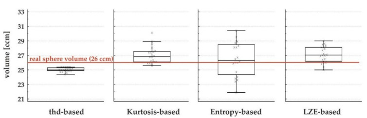
Positron emission tomography (PET) provides important additional information when applied in radiation therapy treatment planning. However, the optimal way to define tumors in PET images is still undetermined. As radiomics features are gaining more and more importance in PET image interpretation as well, we aimed to use textural features for an optimal differentiation between tumoral tissue and surrounding tissue to segment-target lesions based on three textural parameters found to be suitable in previous analysis (Kurtosis, Local Entropy and Long Zone Emphasis). Intended for use in radiation therapy planning, this algorithm was combined with a previously described motion-correction algorithm and validated in phantom data. In addition, feasibility was shown in five patients. The algorithms provided sufficient results for phantom and patient data. The stability of the results was analyzed in 20 consecutive measurements of phantom data. Results for textural feature-based algorithms were slightly worse than those of the threshold-based reference algorithm (mean standard deviation 1.2%-compared to 4.2% to 8.6%) However, the Entropy-based algorithm came the closest to the real volume of the phantom sphere of 6 ccm with a mean measured volume of 26.5 ccm. The threshold-based algorithm found a mean volume of 25.0 ccm. In conclusion, we showed a novel, radiomics-based tumor segmentation algorithm in FDG-PET with promising results in phantom studies concerning recovered lesion volume and reasonable results in stability in consecutive measurements. Segmentation based on Entropy was the most precise in comparison with sphere volume but showed the worst stability in consecutive measurements. Despite these promising results, further studies with larger patient cohorts and histopathological standards need to be performed for further validation of the presented algorithms and their applicability in clinical routines. In addition, their application in other tumor entities needs to be studied.
Keywords: lung cancer; positron emission tomography (PET); radiation therapy treatment planning; radiomics; textural features; tumor volume segmentation.
Conflict of interest statement
R.A.B. is a consultant for Bayer Healthcare (Leverkusen, Germany) and Eisai GmbH (Frankfurt, Germany). R.A.B. has a non-commercial research agreement and is on the speakers list of Mediso Medical Imaging (Budapest, Hungary). M.E. is a consultant for Bayer Healthcare (Leverkusen, Germany) and Eisai GmbH (Frankfurt, Germany), IPSEN and Novartis. All other authors declare that there is no conflict of interest and have given their consent for scientific analysis and publication.
Figures


References
-
- Nestle U., Schimek-Jasch T., Kremp S., Schaefer-Schuler A., Mix M., Küsters A., Tosch M., Hehr T., Eschmann S.M., Bultel Y.-P., et al. Imaging-based target volume reduction in chemoradiotherapy for locally advanced non-small-cell lung cancer (PET-Plan): A multicentre, open-label, randomised, controlled trial. Lancet Oncol. 2020;21:581–592. doi: 10.1016/S1470-2045(20)30013-9. - DOI - PubMed
LinkOut - more resources
Full Text Sources
Research Materials
