. 2022 Feb 26;12(3):597.
doi: 10.3390/diagnostics12030597.

Measurement of the Depth of Lesions on Proximal Surfaces with SWIR Multispectral Transillumination and Reflectance Imaging

Affiliations

Measurement of the Depth of Lesions on Proximal Surfaces with SWIR Multispectral Transillumination and Reflectance Imaging

Yihua Zhu et al. Diagnostics (Basel). .

Abstract

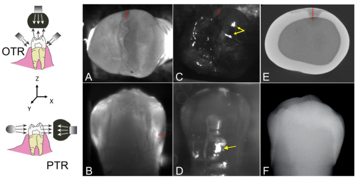
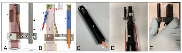
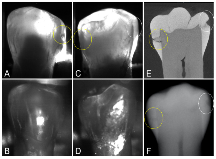
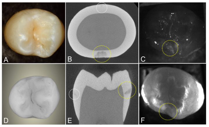
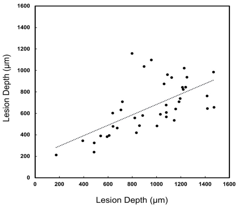
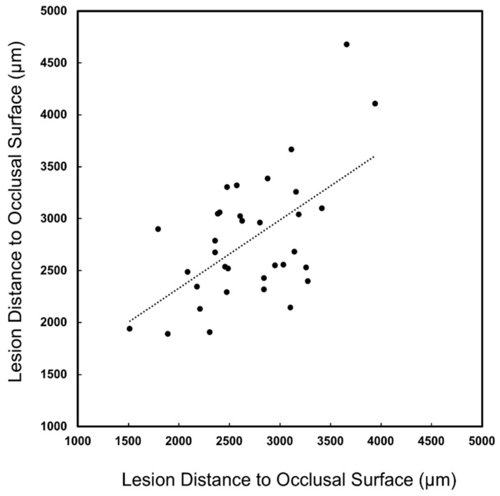
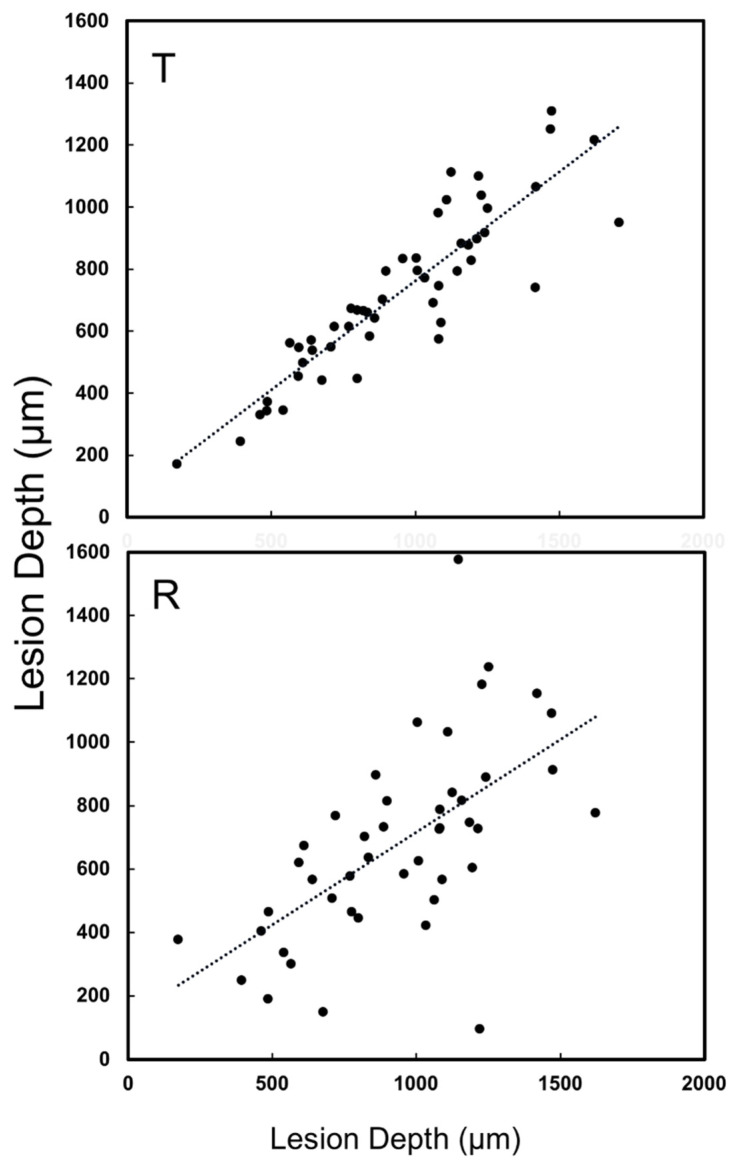
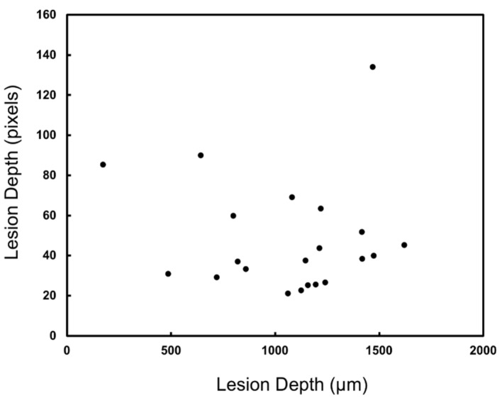
The aim of this study was to compare the diagnostic performance of dual short-wavelength infrared (SWIR) transillumination and reflectance multispectral imaging devices for imaging interproximal lesions with radiography using extracted teeth that had been imaged with micro-computed tomography (microCT). Thirty-six extracted teeth with 67 lesions on the proximal surfaces were imaged using a newly fabricated SWIR multispectral proximal transillumination and reflectance imaging device along with an existing SWIR multispectral occlusal transillumination and reflectance device. The ability of SWIR imaging and radiography to detect lesions and accurately assess lesion dimensions were compared using microCT as a standard. Occlusal and proximal transillumination and occlusal reflectance performed best for imaging interproximal lesions while proximal reflectance was not useful for imaging interproximal lesions from tooth buccal and lingual surfaces. There was high correlation of the lesion dimensions measured in occlusal and proximal transillumination images compared to microCT and moderate correlation in occlusal reflectance images. The correlation between the lesion depth measured in radiographs and the lesion depth measured with microCT was not significant. This study demonstrates that SWIR occlusal and proximal transillumination and SWIR occlusal reflectance images are useful for imaging interproximal lesions and they provide more accurate measurements of lesion severity.

Keywords: SWIR imaging; caries detection; reflectance; transillumination.


Conflict of interest statement

The authors declare no conflict of interest.

Figures

Figure 1
Figure 1
Diagrams of the occlusal (OTR) and proximal (PTR) dual short-wavelength infrared (SWIR) transillumination and reflectance probes used in this study. The orientation of the XYZ axes with respect to the long axis of the tooth is shown. (A) Occlusal transillumination image, (B) proximal transillumination image (buccal surface), (C) occlusal reflectance image and (D) proximal reflectance (buccal surface) image, (E) transverse microCT slice in the XY plane of a tooth, and (F) dental radiograph of a tooth with an interproximal lesion is shown. The red dotted line represents the lesion depth from the proximal surface (X direction), the green dotted line represents the lesion width (Y direction) and the blue dotted line represents the vertical distance of the lesion to the tooth occlusal surface (Z direction). The yellow arrows in the reflectance images (C,D) are areas of specular reflection that can potentially cause false positives.
Figure 2
Figure 2
(A) 3D rendering and (B) cross-section of the dual SWIR proximal probe is shown. Major parts include: (1) probe main body, (2) reflectance backpiece, (3) sliding frame, (4) Delrin rod holder, (5) Delrin tube, (6), right-angled rectangular aluminum reflector, (7) right-angle cylindrical aluminum reflector, (8) Teflon tube for reflectance light source, (9) Teflon tube for proximal transillumination light source, (10) optical fiber delivering 1450 nm light for reflectance, (11) optical fiber delivering 1300 nm light for proximal transillumination. Proximal transillumination light propagation is shown in orange dashed arrows. Reflectance light propagation is shown with red dashed arrows. (C) Proximal transillumination light source (11) is encased in a Teflon tube placed inside a Delrin tube (5). The cylindrical aluminum mirror (7) reflects anisotropically scattered light from the Teflon tube and directs it to the sample. (D) Fully assembled dual SWIR proximal probe for in vitro use. (E) For in vivo use, the sliding frame (3) can be replaced with a soft/flexible holder.
Figure 3
Figure 3
Proximal transillumination and reflectance images of a tooth with two interproximal lesions on opposing proximal surfaces. Lesion 1 (yellow circle) is more severe and penetrates to the dentin while lesion 2 (white circle) is limited to the enamel. (A) Transillumination and (B) Reflectance images with the imager on the buccal side of the tooth. (C) Transillumination and (D) Reflectance with the imager on the lingual side of the tooth. (E) MicroCT slice (F) and Dental X-Radiograph are also shown. Neither lesion is visible in the proximal reflectance images.
Figure 4
Figure 4
Images of a tooth with two interproximal lesions, one that is quite small and is only visible in the microCT images (white circle) and one much larger that was also visible in the SWIR images (yellow circle). (A) color and SWIR (C) occlusal reflectance and (F) occlusal transillumination are shown along with a (D) microCT surface rendering of the tooth and extracted slices (B) transverse and parallel. (E) to the long axis of the tooth.
Figure 5
Figure 5
The lesion depth was measured in proximal transillumination mode (Xpt) and microCT (Xµ). There was a significant correlation between the two methods (p < 0.05) with R2 = 0.40 and 40 of the 67 lesions had sufficient contrast to perform the measurements.
Figure 6
Figure 6
Depth below the occlusal surface measured in transillumination mode (Zot) and microCT (Zµ) There was a significant correlation between the two methods (p < 0.05) with R2 = 0.33 and 34 of the 67 lesions had sufficient contrast to perform the measurements.
Figure 7
Figure 7
(T) Lesion depth measured in occlusal transillumination mode (Xot) and microCT (Xµ). There was a significant correlation between the two methods (p < 0.05) with R2 = 0.80 and 51 out of the 67 lesions had sufficient contrast to perform the measurements. (R) Lesion depth measured in occlusal reflectance mode (Xor) and microCT (Xµ). There was a significant correlation between the two methods (p < 0.05) with R2 = 0.38 and 45 of the 67 lesions had sufficient contrast to perform the measurements.
Figure 8
Figure 8
Lesion depth was measured in radiography (Xr) and microCT (Xµ). There was no significant correlation between the two methods (p > 0.05). Only 22 of the 67 lesions had sufficient contrast to perform the measurements.

Similar articles

Cited by

References

    1. Buhler C., Ngaotheppitak P., Fried D. Imaging of occlusal dental caries (decay) with near-IR light at 1310-nm. Opt. Expr. 2005;13:573–582. doi: 10.1364/OPEX.13.000573. - DOI - PubMed
    1. Staninec M., Lee C., Darling C.L., Fried D. In vivo near-IR imaging of approximal dental decay at 1,310 nm. Lasers Surg. Med. 2010;42:292–298. doi: 10.1002/lsm.20913. - DOI - PMC - PubMed
    1. Jones G., Jones R.S., Fried D. Transillumination of interproximal caries lesions with 830-nm light; Proceedings of the Lasers in Dentistry X, Proc SPIE; San Jose, CA, USA. 28 May 2004; pp. 17–22.
    1. Jones R.S., Huynh G.D., Jones G.C., Fried D. Near-IR Transillumination at 1310-nm for the Imaging of Early Dental Caries. Opt. Expr. 2003;11:2259–2265. doi: 10.1364/OE.11.002259. - DOI - PubMed
    1. Fried D., Featherstone J.D.B., Darling C.L., Jones R.S., Ngaotheppitak P., Buehler C.M. Early Caries Imaging and Monitoring with Near-IR Light. Dent. Clin. N. Am. Incip. Hidden Caries. 2005;49:771–794. doi: 10.1016/j.cden.2005.05.008. - DOI - PubMed