

Local and Metastatic Relapses in a Young Woman with Papillary Squamous Cell Carcinoma of the Uterine Cervix
- PMID: 35328152
- PMCID: PMC8946994
- DOI: 10.3390/diagnostics12030599
Local and Metastatic Relapses in a Young Woman with Papillary Squamous Cell Carcinoma of the Uterine Cervix
Abstract
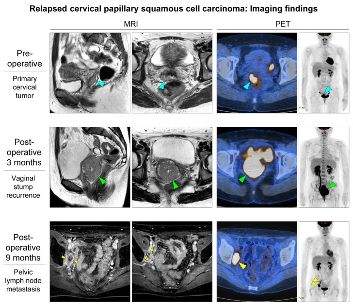
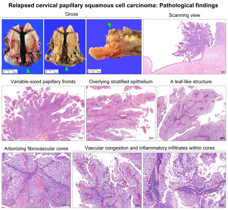
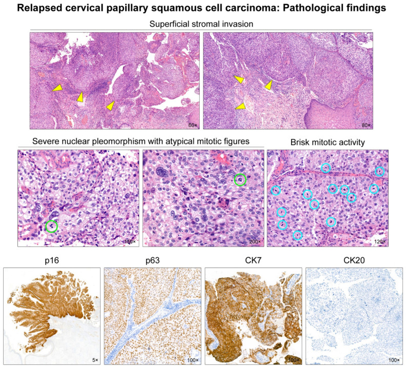
Papillary squamous cell carcinoma (PSCC) is a rare histological type of cervical carcinoma whose biological behavior has not been fully established. A 33-year-old woman with an exophytic cervical mass underwent radical hysterectomy and bilateral pelvic lymph node dissection. Histological examination of the tumor revealed numerous papillary fronds lined by atypical stratified squamous cells, resembling high-grade squamous intraepithelial lesions or urothelium. She was diagnosed with stage IB1 PSCC. Three months postoperatively, a 5.7 cm vaginal stump mass was detected. She received chemoradiotherapy, which helped her achieve a complete response. However, nine months postoperatively, she developed pelvic lymph node metastases. We present a rare case of recurrent cervical PSCC in a young woman. PSCC of the uterine cervix can recur rapidly within just a few months and become aggressive, as in the present case.
Keywords: cervix; metastasis; papillary squamous cell carcinoma; recurrence.
Conflict of interest statement
The authors declare no conflict of interest.
Figures
Similar articles
-
Papillary Squamotransitional Cell Carcinoma of the Uterine Cervix with Atypical Presentation: A Case Report with a Literature Review.Medicina (Kaunas). 2022 Dec 14;58(12):1838. doi: 10.3390/medicina58121838. Medicina (Kaunas). 2022. PMID: 36557040 Free PMC article. Review.
-
Papillary squamous cell carcinoma of the cervix: Two cases and a review of the literature.Gynecol Oncol Rep. 2016 Oct 15;18:18-21. doi: 10.1016/j.gore.2016.10.003. eCollection 2016 Nov. Gynecol Oncol Rep. 2016. PMID: 27790636 Free PMC article.
-
Papillary Squamotransitional Cell Carcinoma of the Uterine Cervix: A Case Report and Review of the Literature.Case Rep Obstet Gynecol. 2016;2016:7107910. doi: 10.1155/2016/7107910. Epub 2016 Aug 30. Case Rep Obstet Gynecol. 2016. PMID: 27656303 Free PMC article.
-
Papillary squamotransitional cell carcinoma of the uterine cervix: A histomorphological and immunohistochemical study of nine cases.Indian J Med Paediatr Oncol. 2013 Apr;34(2):66-71. doi: 10.4103/0971-5851.116177. Indian J Med Paediatr Oncol. 2013. PMID: 24049289 Free PMC article.
-
Papillary squamous cell carcinoma of the uterine cervix: report of a case with HPV 16 DNA and brief review.Gynecol Oncol. 1996 Dec;63(3):388-91. doi: 10.1006/gyno.1996.0341. Gynecol Oncol. 1996. PMID: 8946877 Review.
Cited by
-
Papillary Squamotransitional Cell Carcinoma of the Uterine Cervix with Atypical Presentation: A Case Report with a Literature Review.Medicina (Kaunas). 2022 Dec 14;58(12):1838. doi: 10.3390/medicina58121838. Medicina (Kaunas). 2022. PMID: 36557040 Free PMC article. Review.
-
Clinical Outcomes of Pleomorphic High-grade Squamous Intraepithelial Lesions of the Uterine Cervix: A Single-institutional Experience of 44 Cases.In Vivo. 2024 Nov-Dec;38(6):3050-3058. doi: 10.21873/invivo.13789. In Vivo. 2024. PMID: 39477401 Free PMC article.
-
Squamotransitional Cell Carcinoma of the Uterine Cervix with Ovarian Metastasis and Benign Brenner Tumor: A Case Report.Reports (MDPI). 2023 Nov 13;6(4):54. doi: 10.3390/reports6040054. Reports (MDPI). 2023. PMID: 40729177 Free PMC article.
-
Value of loop electrosurgical excision procedure conization and imaging for the diagnosis of papillary squamous cell carcinoma of the cervix.Front Oncol. 2023 Jul 5;13:1166818. doi: 10.3389/fonc.2023.1166818. eCollection 2023. Front Oncol. 2023. PMID: 37476381 Free PMC article.
-
β-lapachone suppresses carcinogenesis of cervical cancer via interaction with AKT1.Front Pharmacol. 2025 Feb 20;16:1509568. doi: 10.3389/fphar.2025.1509568. eCollection 2025. Front Pharmacol. 2025. PMID: 40051559 Free PMC article.
References
-
- Ren H., Pors J., Chow C., Ta M., Stolnicu S., Soslow R., Huntsman D., Hoang L. Evaluation of human papillomavirus (HPV) prediction using the International Endocervical Adenocarcinoma Criteria and Classification system, compared to p16 immunohistochemistry and HPV RNA in-situ hybridization. J. Pathol. Transl. Med. 2020;54:480–488. doi: 10.4132/jptm.2020.07.18. - DOI - PMC - PubMed
LinkOut - more resources
Full Text Sources
